3D Imaging of Brain Lesions May Spot Those Most Likely to Heal, Guiding Treatment
Written by |
A new diagnostic method for multiple sclerosis (MS) that uses 3D analysis of a patient’s brain may be able to tell physicians which lesions there are more likely to heal with time and which are not, and as such could be a game-changer in treating the disease, according to the researchers who developed the approach.
Their study, “Three‐Dimensional Lesion Phenotyping and Physiologic Characterization Inform Remyelination Ability in Multiple Sclerosis,” was published in the Journal of Neuroimaging.
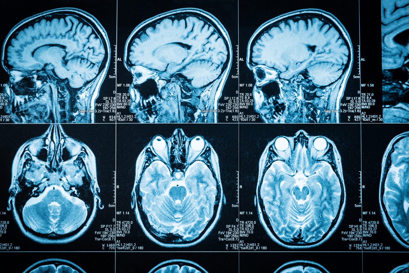
Physicians rely heavily on magnetic resonance imaging (MRI) brain scans to diagnose, monitor, and predict the progression of MS. But current MRI techniques have several limitations, including 2D visualization of brain structures, lack of specificity regarding disease origin, and overly frequent false positive findings of lesions due to the tool’s high sensitivity.
“Three-dimensional [analysis] of MS lesions provides a comprehensive view of their shape and surface characteristics. Such observations [may provide] important information regarding MS diagnosis and disease progression,” the researchers wrote.
Scientists with the Center for BrainHealth at the University of Texas (UT) at Dallas, working with colleagues at UT Southwestern, set out to develop a new diagnostic tool for MS based on 3D imaging analysis of brain lesions visible in MRI brain scans.
They analyzed 109 brain lesions from 23 MS patients. Some lesions, they found, were surrounded by high levels of oxygen — a clear indication that these lesions were metabolically active and more likely to heal over time.
Based on this information, the researchers came up with a noninvasive biomarker — the blood oxygen level dependent (BOLD) slope — that compares oxygen levels on the lesions to those of the surrounding tissues, so to quickly distinguish between metabolically active and inactive lesions.
They then created 3D images of patients’ brain lesions using a new and to-be-patented technology, and found that metabolically active lesions tended to be more spherical and had a rough surface. Metabolically inactive lesions had lower levels of surrounding oxygen, and tended to have more irregular shapes and a smoother surface.
“Such observations could not have been made with a conventional 2-D approach. In addition, the short acquisition time for BOLD slope, and minimal degree of post processing required to calculate these outcomes may further increase its potential in the clinical management of MS patients,” the researchers wrote.
The team also discovered that white matter in metabolically active lesions remained more intact compared to metabolically inactive lesions. (White matter refers to areas of the central nervous system — composed by the brain, brainstem, and cerebellum — made up of myelinated nerve segments, or axons, that are responsible for the transmission of nerve signals.)
“This diagnostic method represents a significant advance in our field, considering the new MS drugs being developed to heal damaged areas of the brain,” Dinesh Sivakolundu, the study’s lead author, and a teaching and doctoral student at the Center for BrainHealth, said in a press release.
“Using our new technology, we could potentially determine which patients would benefit from such new drugs and which patients would not,” Sivakolundu added.
This new 3D imaging technique could provide a “platform for disease surveillance and outcome quantification involving myelin repair therapeutics,” the researchers concluded.