
Immune Treg Cells Seen to Ease Paralysis in Mouse Model of MS
Written by |
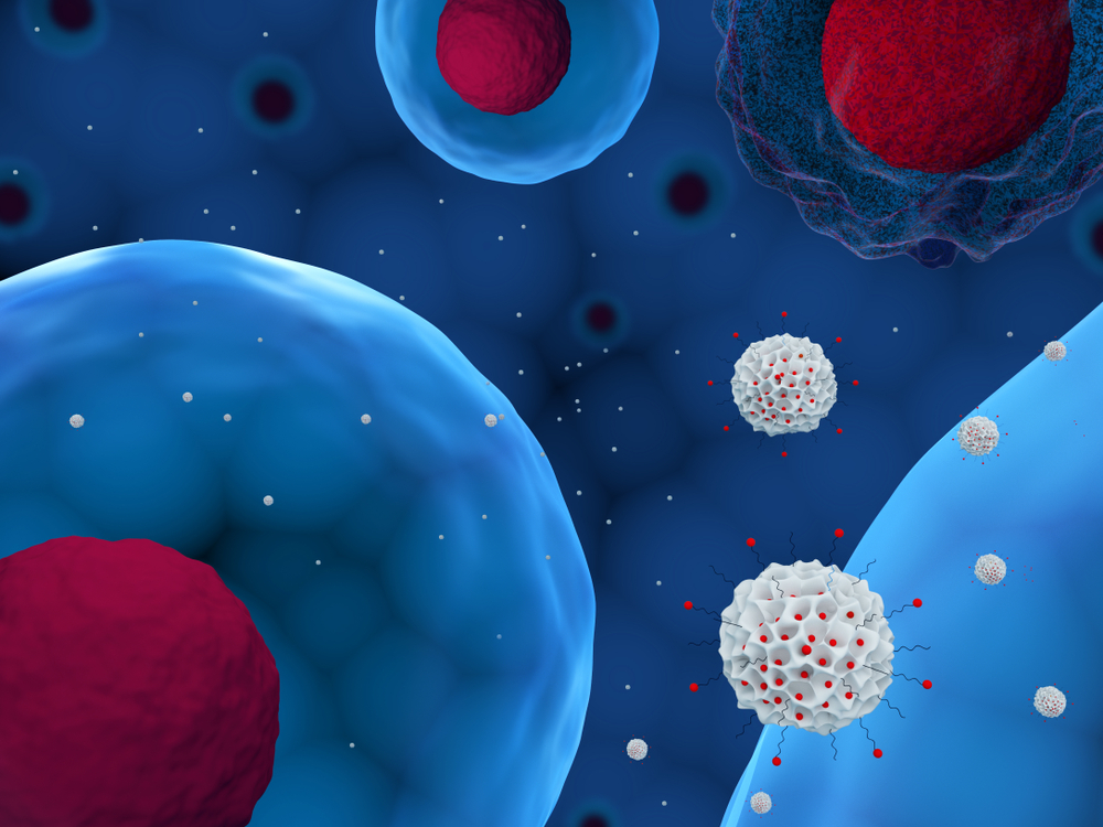
Regulatory T-cells (Tregs) — immune cells that normally dampen immune and inflammatory responses by inhibiting the activity of pro-inflammatory immune cells — enabled mice in a model of multiple sclerosis to partly recover from limb and tail paralysis, scientists reported.
Tregs can do this by preventing a subtype of pro-inflammatory T-helper cells, called Th17, from interacting with another type of immune cell, called antigen-presenting cells (APCs), the team found. Tregs can also block calcium signaling in Th17 cells, further enhancing their ability to counteract Th17’s pro-inflammatory activity.
These findings help in understanding the molecular mechanisms at play in multiple sclerosis (MS), and may lead to new forms of treatment based on the use of T-cells.
The study, “Regulatory T cells suppress Th17 cell Ca2+ signaling in the spinal cord during murine autoimmune neuroinflammation,” was published in the journal PNAS.
“Building on our years of expertise in immunoimaging and calcium signaling, this study highlights Th17 and Treg cell interactions, their motility characteristics, and intracellular signaling, thus providing new insights into the pathophysiology [disease mechanisms] of MS,” Michael Cahalan, PhD, professor and chair of the department of Physiology and Biophysics at the University of California, Irvine (UCI) School of Medicine, and the study’s senior author, said in a press release.
“Our results illustrate how a regulatory T cell-based immunotherapy may be instrumental in limiting demyelination in MS,” Cahalan added.
The interaction between different types of immune cells is known to be vital for controlling the body’s immune and inflammatory responses, but it is still unclear how Tregs position themselves to counter the inflammatory activity of Th17 cells.
To address this and also investigate how Tregs interact with APCs and Th17 cells in the context of inflammation, the researchers used advanced high-resolution imaging to visualize portions of the spinal cord of mice with chronic experimental autoimmune encephalomyelitis (EAE), a disease that mimics MS in humans.
They found that Th17 cells are the first to arrive to the animals’ spinal cord, before the onset of EAE symptoms. Once in the spinal cord, these cells became active by interacting with APCs, and started to show high calcium signaling activity before the arrival of Tregs.
Despite a later appearance, the Tregs remained in the spinal cord during chronic stages of the disease. They surrounded APCs, effectively preventing their interaction with Th17 cells. Tregs were also seen to block calcium signaling in Th17 cells, further limiting the activity of these cells.
“We discovered a unique ‘repetitive scanning motility’ by which Treg cells (the good guys) dampen calcium signaling in pathogenic [disease-causing] Th17 cells (the bad guys), and help to resolve neuroinflammation and limit reactivation of Th17 cells in the spinal cord,” Shivashankar Othy, PhD, the study’s lead author, said.
While Th17 cells outnumbered Tregs in the spinal cord, inflammation persisted and the animals started to experience tail and limb paralysis, the researchers reported. But during the chronic stage of EAE, the numbers of Th17 cells started to drop while those of Tregs remained stable, and the paralysis eased.
“These findings will help to understand how Treg cells prevent autoimmunity and dampen immune responses, and how autoimmune diseases can be effectively targeted using Treg-based cellular therapies,” the investigators wrote.


Leave a comment
Fill in the required fields to post. Your email address will not be published.