
In Creating ‘Immune Cell Atlas of Brain,’ Scientists Find Microglia Highly Complex in Behavior and Role
Written by |

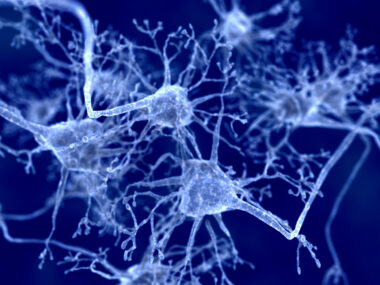
In mapping the immune system of the brain in mice and humans, scientists in Germany found that microglia — a type of nerve cell located in the central nervous system and responsible for supporting and protecting neurons — share the same core design, but behave differently depending on the specific function each are meant to play.
Researchers believe this discovery of relevance to understanding disease mechanisms and identifying potential therapeutic targets in neurodegenerative and neuroinflammatory disorders, including multiple sclerosis (MS), Alzheimer’s disease and autism spectrum disorders (ASDs).
Findings in the study, “Spatial and temporal heterogeneity of mouse and human microglia at single-cell resolution,” were published in the journal Nature.
The brain is a protected from blood that circulates through the body by a highly selective and semipermeable membrane called the blood-brain barrier (BBB). The membrane’s main purpose is to prevent the brain from being invaded by microbes; it allows only nutrients and other small molecules to pass through.
However, this barrier also prevents immune cells in the blood from reaching the brain and the spinal cord. For this reason, the central nervous system (CNS) developed its own immune system in the form of microglia cells. These special nerve cells appear very early during embryonic development, and their main role is to eliminate germs and dead cells from the brain to protect neurons.
Up until now, scientists believed there were different types of microglia in the brain, one for each specific function.
But a team of researchers at the University of Freiburg, working with other scientists, showed that all microglia share the same core signature. However, they behave differently, at different time points and in different regions of the brain, depending on the specific function they are meant to serve.
“We were able to show that there is only a single type of microglia in the brain that exist in multiple flavours,” Marco Prinz, MD, medical director of the Institute of Neuropathology at the Medical Center at the University of Freiburg and corresponding author of the study, said in a press release.
“These immune cells are very versatile all-rounders, not specialists, as has been the textbook opinion up to now,” Prinz added.
Or, as the study noted: “Single-cell analysis of tissues of the central nervous system during homeostasis in mice revealed specific time- and region-dependent subtypes of microglia. Demyelinating and neurodegenerative diseases evoked context-dependent subtypes of microglia with distinct molecular hallmarks and diverse cellular kinetics,” with “corresponding clusters of microglia … identified in healthy human brains, and the brains of patients with multiple sclerosis.”
These findings were based on observations of single cells, made possible by high-resolution microscopy, in different regions and developmental stages of the brain of mice and human tissue samples.
Microglia that don’t behave as they should (are dysregulated) have been implicated in multiple neurological disorders, including MS. For this reason, the researchers believe their work may help in better understanding the cellular mechanisms involved in disease onset and progression, as well as in developing new therapies.
“We now possess the first high-resolution immune cell atlas of the human brain. This also enables us to understand how these cells change during course of diseases like MS,” Prinz said.
“In MS patients, we managed to characterize microglia in a state that is specific for multiple sclerosis. We hope that it will be possible in the future to target microglia subsets in [a] harmful state,” he concluded.

Leave a comment
Fill in the required fields to post. Your email address will not be published.