Fact-checked by
Multiple sclerosis lesions
Multiple sclerosis (MS) is a neurological disorder caused by the body’s immune system mistakenly attacking the myelin sheath, a fatty coating that surrounds nerve fibers. The resulting nerve damage, which can be visualized as lesions or areas of abnormal tissue in the nervous system, ultimately causes the symptoms of MS.
What is an MS lesion?
An MS lesion, sometimes also called a plaque or scar, is an area in the central nervous system that has been damaged by the immune system’s attack. Lesions are thought to occur upon a backdrop of inflammation. The size and shape of MS lesions can vary widely, and they can affect different regions of the central nervous system. MS lesions can form in the brain, spinal cord, and optic nerves.
Within a lesion, there is a loss of myelin — a process referred to as demyelination — that often is accompanied by scarring, or sclerosis. This scarring is actually what gives MS its name, as the disease is characterized by “multiple” instances of “sclerosis” in the central nervous system.
Picture of MS lesions
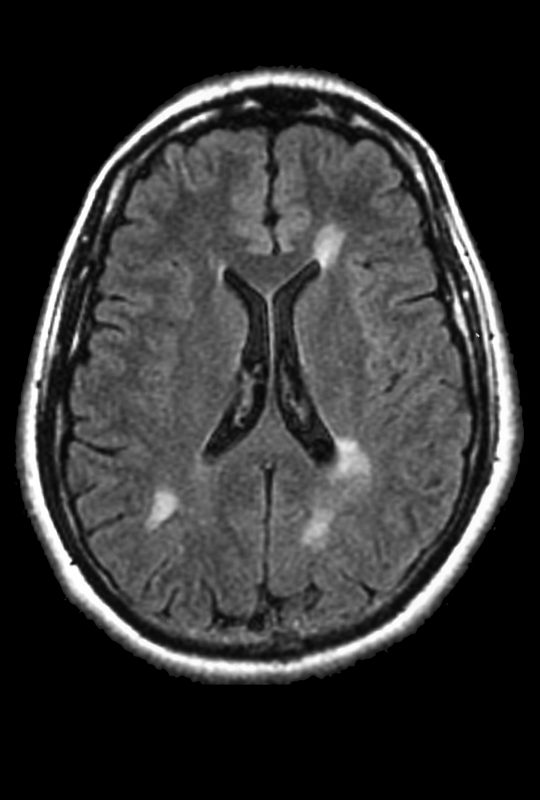
Shown is an image from a brain MRI of a young multiple sclerosis patient. The white spots correspond to demyelinating lesions. (Credit: ISM/SOVEREIGN)
Can you have MS without lesions?
MS is defined by the presence of lesions in the central nervous system. The indicators used for making an early diagnosis are called the McDonald criteria. Under these formal guidelines, a person must show evidence of lesions that accrue over time and that affect at least two of four regions in the central nervous system — three areas of the brain, and the spinal cord. The brain areas affected in MS are the periventricular, juxtacortical or cortical, and infratentorial. If a person does not meet these criteria, then the clinical case cannot be considered MS.
Usually, MS lesions are detected via imaging using MRI scans. It is possible for a person to have MS lesions that aren’t visible on these scans — for example, some lesions may be too small to be seen. Under the current McDonald criteria, symptoms that are indicative of an MS lesion in a particular region of the nervous system may be used as evidence for a lesion in that region, but it is crucial to rule out other possible explanations for symptoms in such cases.
Active and inactive lesions
A so-called active lesion represents an area of ongoing inflammation in which the immune system is currently attacking the myelin sheath. By contrast, an inactive lesion is a spot where there is myelin loss (demyelination) and nerve damage as a result of an earlier immune attack, but there is no longer ongoing or active inflammation at the site of the lesion.
Some lesions show evidence of sustained inflammatory activity over time, especially in progressive types of MS. A chronically active lesion is sometimes called a “smoldering” lesion — other terms are “paramagnetic rim” or “iron rim” lesions, as the ongoing inflammation is characterized by a “rim” of iron-containing immune cells around the lesion site.
How are lesions detected in MS?
MRI
The gold standard for detecting and monitoring MS lesions in the brain and spinal cord is magnetic resonance imaging, commonly known as MRI. MRI is a non-invasive imaging technique that’s often used to determine the number and volume of lesions in a given individual, a parameter referred to as the “lesion load.”
In simple terms, MRI works by using powerful magnetic fields and computer-generated radio waves as a means to create detailed images of tissues and organs. Unlike other types of scans, the MRI technique does not expose the patient to radiation.
Several types of MRI scans can be useful for detecting specific forms of MS lesions. The ones most commonly used in clinical practice include T1-weighted scans that detect active inflammatory lesions, and T2-weighted scans, which detect both old and active lesions (the total lesion load).
Can MS lesions be mistaken for migraine lesions?
Migraine is a condition defined by periodic, severe headaches typically characterized by a throbbing or pulsing pain, nausea, and extreme sensitivity to lights and sounds. There are some similarities between migraine and MS, as both are chronic conditions affecting the nervous system, and both tend to be characterized by relapses or attacks — times in which symptoms suddenly worsen.
People with migraine may have lesions in their brains resulting from white matter abnormalities, and these may appear similar to MS lesions on MRI scans. Thus, it can be difficult to distinguish between migraine and MS lesions, and there are many reports of people with migraine who initially received an incorrect diagnosis of a form of MS, or vice versa.
Further complicating matters, while headaches are not considered a typical symptom of MS, migraine and MS can co-occur — in fact, research suggests that rates of migraine are two to three times higher among MS patients than in the general population. A review study published in 2020 estimated that roughly 30% of MS patients experience migraines.
More like this...


Living with MS
Symptoms of MS vision problems
Living with MS
High-dose vitamin D delays MS progression
According to the American Migraine Foundation, the MRI abnormalities seen in some patients may not even be considered “lesions” as they do not cause clear damage or disease-related signs; it is believed that these abnormalities are generally not associated with any type of neurological issues or an increased risk of cognitive decline.
Symptoms of MS lesions
How do lesions correlate with MS symptoms?
A lesion can interfere with the normal function of nerve cells, blocking or slowing normal electrical communication or nerve impulses in the brain and spinal cord. This can directly contribute to MS symptoms, with the specific effect depending on the location of the lesion.
However, not all lesions cause obvious symptoms. Sometimes patients can develop new lesions without associated disease manifestations — these lesions are referred to as “clinically silent.” Conversely, patients may also have symptoms in the absence of a clearly detectable lesion in a corresponding part of the central nervous system.
Furthermore, it is not always possible to make a direct correlation between the location and number of lesions and the clinical signs and symptoms a patient experiences as part of disease progression.
Generally, according to the National MS Society, lesions in areas such as the spinal cord or optic nerve are likely to result in disease manifestations.
What do MS lesions feel like?
Even though the central nervous system is packed with nerve cells, the brain tissue itself does not have so-called noniceptors — the sensory nerve fibers that detect pain and potentially damaging stimuli. Thus, MS lesions themselves cannot be felt. Instead, they may induce symptoms that result from tissue damage to the nervous system.
The symptoms and signs of MS can vary depending on the particular location of the lesion. For example, a lesion in the optic nerves, which connect the eyes to the brain, can cause vision problems. Meanwhile, lesions in the spinal cord can cause unusual sensations — such as tingling or numbness — or motor symptoms, including loss of balance and/or coordination. Spinal cord lesions also may be associated with bladder and bowel impairments.
Lesions in the brainstem and cerebellum, toward the base of the brain, may cause symptoms that affect the face, including weakness, unusual sensation, double vision, and difficulty swallowing. Damage occurring toward the top of the brain, specifically the cortex and cerebrum, often does not cause obvious symptoms, though lesions in those areas may be associated with cognitive challenges and depression.
These associations are far from absolute, and it is possible for an individual to have MS lesions without any obvious accompanying symptoms, or to have symptoms even though a lesion cannot be clearly visualized on MRI scans. MS patients should talk with their healthcare providers whenever they experience any new symptoms.
Scroll horizontally to view all columns -->
| Lesion location | Possible associated symptoms |
|---|---|
| Spinal cord |
|
| Optic nerves |
|
| Lower back of the brain (infratentorial region, including the cerebellum and brainstem) |
|
| Central section of the brain (periventricular region, including the frontal horns, posterior horns, and ventricular bodies) |
|
| Outermost section of the brain (cortical or juxtacortical regions, including the cortex and cerebrum) |
|
Can MS lesions shrink and go away?
Over time, the number and size of lesions in one person may increase, decrease, or remain stable. Generally, an increase in the number or size of lesions, or the appearance of lesions with new inflammatory activity, is indicative of worsening disease.
The repair and regeneration of myelin in MS lesions are, to some extent, possible. Cells called oligodendrocyte precursor cells are recruited to the lesion site, where they differentiate into mature oligodendrocytes. These mature cells are able to produce myelin and can create a new sheath for nerve fibers, a process referred to as remyelination. Thus, sometimes, lesions can be repaired and disappear, and not be detected on subsequent MRI scans.
Does MS affect your skin and bones?
MS attacks the myelin sheath in nerve cells of the central nervous system, comprising the brain, spinal cord, and optic nerves. This damage is seen as lesions through imaging techniques, namely MRI. This type of MRI-detected lesions should not be confused with lesions in skin and/or bones, some of which can be seen by the naked eye, that occur in other medical conditions.
Nonetheless, MS can affect a patient’s skin by causing abnormal sensations such as itching, numbness, or tingling. These sensations are a result of damage to nerves, not due to a direct effect on the skin.
There also has been increasing evidence that MS is associated with an increased risk of osteopenia, or the loss of bone density, and osteoporosis — a condition characterized by weak bones that are more prone to fractures. The reasons for this are linked to factors known to reduce bone mass, including the use of corticosteroids, a standard treatment for acute MS relapses, vitamin D deficiency, and the prolonged motor disability of MS patients. Long-term motor disability reduces the mechanical loading of bone.
Multiple Sclerosis News Today is strictly a news and information website about the disease. It does not provide medical advice, diagnosis, or treatment. This content is not intended to be a substitute for professional medical advice, diagnosis, or treatment. Always seek the advice of your physician or other qualified health provider with any questions you may have regarding a medical condition. Never disregard professional medical advice or delay in seeking it because of something you have read on this website.
FAQs about MS lesions
MS lesions are dynamic and can change as time goes on. Over time, an individual lesion or area of abnormal tissue may remain the same size, it may grow, or it may shrink or disappear entirely.
While MRI scans are the gold standard for imaging MS lesions, some lesions may not always be clearly visible on scans. For example, some lesions may be too small to reliably be seen using MRI.
Unlike an MRI, which uses powerful magnetic fields to image the body’s internal structures, a computed tomography (CT) scan images the body using a series of X-ray images taken from different angles. While it is possible for MS lesions to be visible on a CT scan, these scans provide less detail than MRI.
There is no solid answer at this point. Some research suggests that MS lesions are more likely to develop after periods of heightened stress. Further, some studies have reported that stress-reducing interventions may reduce the risk of developing new lesions. Nonetheless, other research has found no such connection, and the potential association between stress and MS lesions — and MS progression in general — remains unproven.
To be diagnosed with multiple sclerosis, a person must show evidence of MS-like lesions that affect multiple areas of the nervous system and develop over time. Although lesions are necessary to confirm a diagnosis of MS, other medical conditions also may result in the development of lesions in the brain. Thus, it is important to rule out other potential causes when determining an MS diagnosis.