Evoked potential test for MS diagnosis
 Reviewed by
An evoked potential (EP) test for multiple sclerosis (MS) can help clinicians assess nerve damage when diagnosing the disease.
MS occurs when the immune system mistakenly attacks myelin, the protective covering around nerve fibers. An MS diagnosis often requires several tests, such as MRI scans to image lesions in the brain and a lumbar puncture, or spinal tap, to analyze cerebrospinal fluid for certain proteins.
EP tests can identify slowed signals that may suggest myelin damage along sensory pathways. Different types evaluate different sensory pathways.
What is an evoked potential test for MS?
EP tests measure the electrical activity of the brain and spinal cord in response to images, sounds, or mild electrical stimulation.
A healthcare specialist, such as a neurologist or neurodiagnostic technologist, places small metal disks called electrodes on the scalp or body before delivering the stimulus. The electrodes measure the response of the brain and central nervous system to the stimuli.
Electrophysiological responses in the brain are electrical signals produced in response to sensory stimuli. They travel faster along healthy nerve pathways.
EP tests measure how long electrical signals take to travel along nerve pathways. They can sometimes detect changes caused by nerve damage even before a person notices symptoms or a neurological examination shows a problem.
The three main types of EP tests used in MS
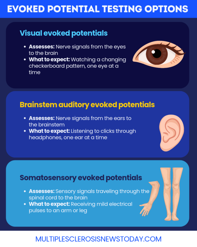
Three main types of EP tests evaluate visual, auditory, and sensory pathways:
- Visual evoked potentials (VEP)
- Brain stem auditory evoked potentials (BAER)
- Somatosensory evoked potentials (SEP)
Visual evoked potentials
A VEP test for MS measures how well electrical signals travel from the eyes through the optic nerves to the part of the brain that processes vision. It can help detect optic nerve damage, including damage associated with optic neuritis.
Physicians often use VEP in an MS diagnosis, and it is the only type of EP test listed in the diagnostic criteria. It can reveal evidence of nerve sheath damage (demyelination) that has not caused noticeable symptoms.
Brain stem auditory evoked potentials
BAER assesses how sound signals travel from the inner ear to the brainstem via the auditory nerve. Slowed responses may suggest damage along these pathways.
Although BAEP is not routinely used to diagnose MS, research suggests it may help assess brainstem dysfunction in people with the disease.
Somatosensory evoked potentials
SEP tracks nerve signals from the nerves outside the brain and spinal cord, called the peripheral nerves, through the spinal cord and into the brain.
Abnormal results may suggest damage along sensory pathways, which can be associated with numbness, reduced sensation, or tingling.
SEP for MS can help assess how well sensory pathways through the spinal cord and brain are functioning.
How is an evoked potential test performed?
EP tests are generally safe and are usually performed in a doctor’s office or hospital. Most people experience little or no discomfort, although some types of testing may cause brief discomfort. Testing often takes 60 to 90 minutes, depending on the tests performed. All EP tests use electrodes to record electrical responses to specific sensory stimulation.
Electrode setup and preparation
Before the appointment, patients should wash their hair and avoid sprays, styling gels, and oils, which can interfere with electrode function.
Individuals having a VEP test should bring any glasses they use, including separate pairs for distance and close work. People can generally eat, drink, and take their usual medications before testing unless their healthcare team gives different instructions.
Electrodes are attached to specific areas of the scalp and, depending on the test type, other locations such as the earlobes, wrists, or the back of the knees.
The equipment records repeated responses to the same stimulus and averages them to produce a clearer pattern. A specialist reviews the timing and shape of these responses.
Procedure by test type
The table below compares electrode placement and the procedure for each test.
Scroll horizontally to view all columns -->
| Test type | Electrode placement | Procedure | What to expect |
|---|---|---|---|
| Visual evoked potentials (VEP) for MS | Scalp | Watching a changing checkerboard pattern on a screen with one eye covered, then repeating with the other eye | Focusing on the screen while electrodes record the brain’s response |
| Somatosensory evoked potentials (SEP) for MS | Scalp and other areas, such as the wrist or back of the knee | Receiving mild electrical pulses to a nerve in an arm or leg while electrodes record how quickly signals reach the brain | Brief discomfort or muscle twitching from the pulses |
| Brainstem auditory evoked response (BAER) for MS | Scalp and earlobes | Listening through headphones to clicking sounds, with each ear tested separately | Sitting quietly while electrodes record responses to the sounds |
Interpreting the results
A specialist, such as a neurologist or neurophysiologist, reviews the specific waveforms and timing of the brain’s responses. They will analyze these patterns for abnormal activity, put together a report, and share the findings with the healthcare team to determine whether the patient would benefit from further testing.
Role of EP tests in MS diagnostic criteria
The diagnostic criteria for MS are known as the McDonald criteria. Of the three types of EP tests discussed, VEP is the one included in the revised MS diagnostic criteria. It can help identify optic nerve damage that may support an MS diagnosis, but it cannot confirm MS on its own.
During an MS diagnosis, neurologists may look for optic neuritis, or inflammation of the optic nerve. VEPs can reveal slowed signaling that suggests optic nerve damage, even without noticeable vision symptoms. These findings must be interpreted alongside other test results.
The 2024 revisions to the McDonald criteria, published in September 2025, recognize the optic nerve as a fifth location where MS-related damage can help establish a diagnosis. Depending on the clinical situation, VEPs, optical coherence tomography (OCT), or MRI may provide evidence of optic nerve involvement.
EP tests cannot confirm MS on their own. Clinicians interpret the results alongside the patient’s medical history, neurological examination, and other tests, which may include MRI scans, spinal fluid analysis, and blood tests to rule out other condtions.
Multiple Sclerosis News Today is strictly a news and information website about the disease. It does not provide medical advice, diagnosis, or treatment. This content is not intended to be a substitute for professional medical advice, diagnosis, or treatment. Always seek the advice of your physician or other qualified health provider with any questions you may have regarding a medical condition. Never disregard professional medical advice or delay in seeking it because of something you have read on this website.