
Estrogen-based Therapy Shows Potential to Protect Vision in Mouse Model
Written by |

An estrogen-based therapy, called indazole chloride (IndCl), was shown to protect against optic nerve damage in a mouse model of multiple sclerosis (MS), a study reported.
IndCl may help to improve vision in people with the disease, but early treatment could be essential, its researchers suggested.
“Our study shows the optic nerve and optic tract, which undergo significant inflammation, demyelination, and axonal damage, are able to restore some function with IndCl treatment with successful attenuation in inflammation and an increase in remyelination,” Seema Tiwari-Woodruff, PhD, the study’s lead author, said in a press release.
The study, “Alleviation of extensive visual pathway dysfunction by a remyelinating drug in a chronic mouse model of multiple sclerosis,” was published in the journal Brain Pathology.
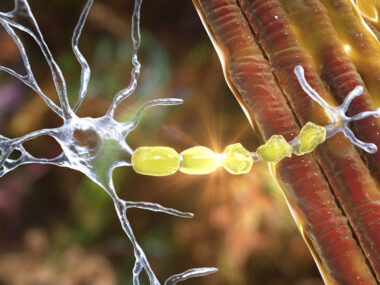
MS is caused by the immune system attacking the myelin sheath, the protective coat around nerve fibers (axons). The optic nerve, which transfers visual information from the retina to the brain via electrical impulses, is heavily myelinated. As a result, up to 50% of people with MS experience optic neuritis — inflammatory demyelination (myelin loss) of the optic nerve — before showing other symptoms.
Most visual studies in MS patients and mouse models focus on the retina and optic nerve itself; however, pathways up to the visual cortex at the back of the brain are also affected. An analysis of the entire visual system is needed.
Additionally, studies have demonstrated the neuroprotective effects of estrogen‐based treatments in reducing inflammatory lesions in MS mouse models. While there are significant side effects associated with such estrogen treatments, including a risk of breast and uterine cancer, these side effects are mediated primarily through estrogen receptor-alpha but not the estrogen receptor-beta.
IndCl selectively stimulates the estrogen receptor-beta, and has demonstrated remyelinating and neuroprotective effects in spinal cord and other axons.
Tiwari-Woodruff and others researchers mostly at the University of California, Riverside, tested the therapeutic potential of IndCl on the entire visual system in a mouse model of MS — the experimental autoimmune encephalomyelitis (EAE) model.
“IndCl has been previously shown in mice to reduce motor disability, increase myelination, and neuroprotection in the spinal cord and corpus callosum,” Tiwari-Woodruff said. “Its effects in the visual system, however, were not evaluated until now.”
MS-like disease was induced in 8- to 12-week-old mice, with EAE disease onset occurring between 10 to 13 days after induction. Peak disease severity was reported between days 15 and 21, and severity maintained through late disease until day 60.
Once mice reach peak disease severity, daily injections of IndCl were administered to a group of these mice, while others served untreated controls.
An analysis of mice retinas found fewer retinal cells, called retinal ganglion cells (RGCs), in the EAE mice compared to healthy mice, but a lesser loss of these cells in EAE mice given IndCl treatment compared to diseased mice left untreated.
“Overall, EAE + IndCl mice demonstrated increased RGC survival compared to EAE + Vehicle [placebo] mice,” the researchers wrote.
Demyelination was assessed using optic nerve sections, which were stained for the myelin basic protein (MBP), a protein that plays a critical role in the organization of myelin sheaths.
Samples from normal mice showed robust MBP staining. In contrast, significant decreases in myelin intensity were observed on EAE mice. While mice treated with IndCl mice showed enhanced MBP staining with fewer demyelinated lesions compared to EAE mice, there remained persistent axon damage.
Normal mice showed a robust population of oligodendrocytes — cells that produce the myelin sheath in axons — compared to EAE mice. IndCl mice showed a significant recovery in oligodendrocyte numbers compared with untreated EAE animals.
Similarly, normal mice had low populations of immune cells, whereas EAE mice had high cell counts, indicative of inflammation. IndCl-treated mice showed a significant decrease in immune cells.
Electron microscopy analysis of optic nerve cross‐sections showed that normal mice had mostly myelinated axons, while EAE mice had an increase in the number of demyelinated axons. IndCl treatment during peak disease significantly lowered the percentage of non-myelinated axons.
“However, many of the axon and myelin pathologies persisted,” the researchers wrote.
In RGC axons along the optic tract leading to the visual cortex, a significant decrease in myelin basic protein staining was observed in EAE mice compared to optic tract sections of healthy mice. This decrease, again, was seen to be weaker in EAE mice treated with IndCl.
Next, the team assessed axon myelination and neuronal changes in the visual cortex, a major visual processing center in humans and mice that is highly myelinated. Again, EAE mice showed lesser MBP staining compared with normal mice, which was fully reversed with IndCl treatment.
Finally, functional effects of visual pathway pathology were measured by recording visually evoked potentials (VEPs). These are electrical signals initiated by brief visual stimuli, using electrodes resting on the skull above the visual cortex.
Healthy mice had a typical VEP pattern, whereas those in the EAE model showed a significant 50% decrease in amplitudes and an increased signal delay (latency). In IndCl-treated mice, the signal delay was significantly lesser compared with untreated EAE mice.
Taken together, these “findings demonstrate the dynamics of visual pathway dysfunction and disability during EAE, along with the importance of early treatment to mitigate EAE‐induced axon damage,” the researchers wrote.
Early treatment is important to maximize the recovery of visual function upon damage.
“Therapeutics [for MS] must target remyelination and prevent further axonal degeneration and neuronal loss,” Tiwari-Woodruff said.
“We treated the MS mice with IndCl at peak disease. If the brain is highly diseased, some of the axons that could potentially restore visual function are too damaged and will not recover. There’s a point of no return,” she added.
“Our paper stresses that to acquire vision improvement, treatment must start early. Early treatment can recover 75%–80% of the original function,” Tiwari-Woodruff concluded.


Leave a comment
Fill in the required fields to post. Your email address will not be published.