New imaging tool detects damaged myelin across large brain areas
Study: It could help determine how well patients respond to medications
Written by |

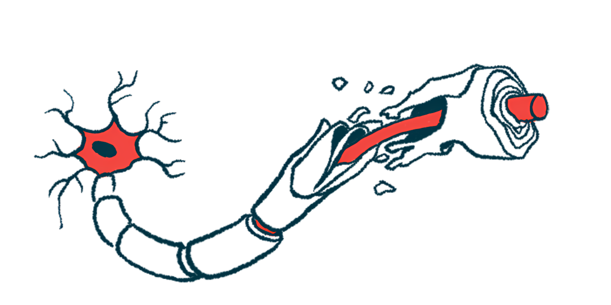
Researchers have developed a highly sensitive imaging technique that can detect damage to myelin, the fatty wrapping around nerve fibers that is damaged in multiple sclerosis (MS), in large areas of the brain, according to a study.
The tool may help assess the extent of myelin damage in people with MS and other demyelinating conditions, and determine how well patients respond to medications designed to boost myelin repair.
“This can guide development and testing of therapies that protect or restore neural wiring,” Tara L. Moore, PhD, co-author of the study at Boston University, said in a university news story. “It may help research into stroke and ischemic injury, chronic traumatic encephalopathy (CTE), multiple sclerosis, Alzheimer’s disease, and other neurodegenerative conditions with myelin involvement, and even age-related cognitive decline.”
The new microscopy imaging method was described in “Birefringence microscopy enables rapid, label-free quantification of myelin debris following induced cortical injury,” which was published in the journal Neurophotonics.
New tool has notable advantages relative to electron microscopy
The myelin sheath is a fatty covering that wraps around nerve fibers, helping them send electrical signals, similar to rubber insulation around a copper wire. In MS, a misguided inflammatory attack damages the myelin sheath in the brain and spinal cord, disrupting normal nerve signaling and ultimately giving rise to MS symptoms.
In scientific studies, the gold standard for assessing myelin integrity is to collect a sample of brain tissue and look at it with an electron microscope. This is an immensely powerful microscope that can visualize structures smaller than a nanometer, or one billionth of a meter, allowing researchers to directly look at myelin in search of cracks and other signs of damage.
However, electron microscopy has some notable limitations. For one thing, preparing samples for electron microscopy is a complex and time-consuming process. For another, electron microscopes can only look at very small areas at a time — smaller than the diameter of a human hair — so they can’t efficiently analyze myelin damage across large brain regions.
A major advantage of BRM over conventional imaging methods is its ability to rapidly image large areas at high resolution without special staining, making it uniquely suited for studying widespread myelin pathology.
In this study, researchers developed a technique to analyze myelin damage using a different type of microscopy called birefringence microscopy, or BRM. Very simply, BRM images tiny structures by shining a light at the sample, then tracking how the light diffracts through it.
BRM has notable advantages relative to electron microscopy. For instance, the techniques for sample preparation are less intensive, and BRM can look at much wider areas, on the order of several square centimeters.
“A major advantage of BRM over conventional imaging methods is its ability to rapidly image large areas at high resolution without special staining, making it uniquely suited for studying widespread myelin pathology,” said Alex Gray, PhD, co-author of the study at Boston University.
BRM-based method detected signs of damaged myelin in monkeys
Gray and colleagues developed their technique by first using standard staining techniques to look for myelin damage in samples of brain tissue from monkeys. They then used machine learning, a form of artificial intelligence, to train a computer on how to detect myelin damage in BRM images of these samples.
The researchers demonstrated that their BRM-based method could effectively detect signs of myelin damage in monkeys that had undergone surgery to induce brain injury. The scientists also showed that their method could detect signs of myelin repair a few weeks after the brain injury occurred.
“These results demonstrate that BRM can enable large-scale investigations into the spatial distribution and extent of myelin damage,” the researchers wrote.
The team noted that their experiments focused specifically on the corpus callosum, a large structure in the center of the brain, in one specific model of monkey brain damage.
Further work will be needed to expand this technique for broader applications. Still, the researchers are hopeful this new technique will help accelerate research in MS and other myelin-related disorders.
“With appropriate characterization and training, this technique can be adapted to other brain regions and disease models, enabling high-throughput, quantitative analysis of myelin structure across conditions, increasing sample sizes, and supporting broader investigations,” the scientists wrote.

