Gut cells prime T-cells to trigger brain inflammation in MS: Study
Results point to gut as potential target site for therapies
Written by |
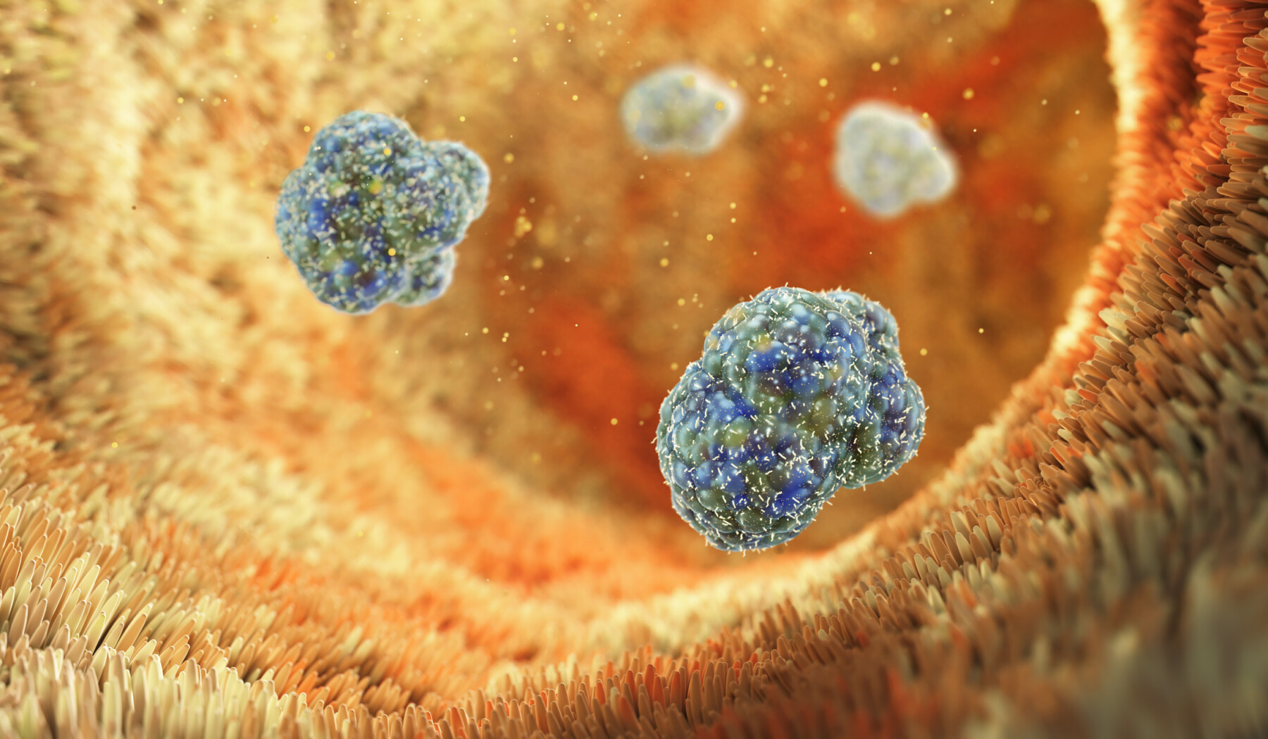
An image shows the intestinal cells lining the gut. (Image from iStock)
- Intestinal epithelial cells activate Th17 cells, driving brain and spinal cord inflammation in MS.
- This gut-brain axis mechanism is supported by findings in MS patients and mouse models.
- Targeting gut immune activation or intestinal epithelial cells may inform new treatments.
The cells that line the inside of the gut — known as intestinal epithelial cells — may play a key role in triggering inflammation that drives multiple sclerosis (MS), a study showed.
The study found that these cells can activate a type of T-cell known as Th17 cells. These pro-inflammatory immune cells then travel to the brain and spinal cord to induce inflammation. Blocking this gut-based immune activation decreased disease severity in a lab model of MS, suggesting this immune pathway may be a valuable therapeutic target in MS.
“While current therapies for MS often target B cells, our study highlights the gut as an important therapeutic site,” Shohei Suzuki, MD, PhD, first author of the study and assistant professor at Keio University in Japan, said in a university press release.
The study, “Intestinal epithelial MHC class II induces encephalitogenic CD4 T cells and initiates central nervous system autoimmunity,” was published in Science Immunology.
MS is marked by inflammation in the brain and spinal cord, collectively known as the central nervous system. The underlying causes of MS are incompletely understood, but a growing body of evidence suggests that disruptions in the composition of gut microbes (known as the gut microbiota) may play a role in MS.

Investigating immune responses in the gut
“Increasing evidence shows that the gut microbiota influences neurological diseases such as Parkinson’s, Alzheimer’s, and MS,” said Tomohisa Sujino, PhD, associate professor at Keio University and the study’s lead author. “However, the mechanisms linking gut microbes, intestinal immunity, and brain inflammation remain unclear. We were keen to identify how gut immune responses contribute to neuroinflammatory diseases.”
Using mice with experimental autoimmune encephalomyelitis (EAE), a lab-induced inflammatory disease used to model MS, the researchers found that Th17 cells accumulate along the intestines. These inflammatory immune cells have previously been linked to CNS inflammation in MS.
Immune cells such as Th17 cells can recognize and attack specific molecular targets, known as antigens. To help guide this process, other cells in the body undertake a process known as antigen presentation, in which they capture pieces of molecules and display them on their surface, almost like putting them in a molecular display case for immune cells to inspect. This system allows the immune system to patrol the body and detect any threats.
Intestinal epithelial cells aren’t usually involved in antigen presentation. But the researchers found that in EAE mice, these gut cells increase the production of major histocompatibility complex class II (MHC II), one of the main molecular display cases used for antigen presentation.
In subsequent experiments, the researchers found that deleting MHC II from intestinal epithelial cells reduced the number of inflammatory Th17 cells, accompanied by a reduction in overall disease severity.
Further tests demonstrated that MHC II on intestinal epithelial cells can trigger Th17 cells to go after antigens found in the brain, and that these inflammatory Th17 cells are able to migrate from the gut into the central nervous system and trigger EAE symptoms.
Collectively, the data support a model in which intestinal cells prime inflammatory immune cells, which then travel to the CNS to drive inflammation. Supporting their work in mouse models, the scientists found that analyses of gut biopsies from MS patients showed elevated levels of Th17 cells and increased MHC II expression in intestinal epithelial cells.
“These findings uncover a conserved gut–central nervous system axis in autoimmunity and position epithelial antigen presentation as a key initiator of neuroinflammation,” the researchers said.
The scientists speculated that this newly uncovered biological pathway may be a key target for new MS treatments.
“Modulating the intestinal microbiota or antigen-presenting activity of [intestinal epithelial cells] represents new approaches to treating autoimmune neurological diseases,” Suzuki said.