
FDA Approves Swedish Company’s Technology for Getting More out of MRI Scans
Written by |
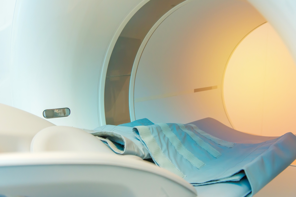
The U.S. Food and Drug Administration has approved technology that the Swedish company SyntheticMR developed to give doctors more information from magnetic resonance imaging scans.
This means the company can begin selling its SyMRI NEURO packages to American medical facilities.
Traditional MRIs offer only one level of contrast when depicting tissue. SyMRI NEURO is the first to offer several levels, giving doctors more information about tissue properties.
This feature can help doctors who are studying multiple sclerosis patients’ brain scans get a better handle on damage to the myelin sheath that protects nerve cells. Deterioration of the sheath is a hallmark of MS.
The additional information should help doctors make better decisions on the diagnosis and treatment of MS patients, SyntheticMR said.
“FDA’s message is a milestone in the history of SyntheticMR,” Stefan Tell, the company’s CEO, said in a press release. “The clearance [approval] means we can now establish our entire product package on the U.S. market and offer clinicians objective decision support for a faster and more reliable diagnosis of their patients.
“A number of studies published this past year show the clinical benefits of SyMRI NEURO,” Tell said. “We are convinced that this is the beginning of a paradigm shift within MRI.”
SyntheticMR developed SyMRI NEURO in collaboration with Juntendo University Hospital researchers in Japan. The partners announced the technology’s development in 2015.
MRI is the preferred imaging tool for diagnosing and tracking MS. Key reasons are that it is the most non-invasive and sensitive method available for looking at the brain, spinal cord and other areas.
It uses magnetic field and radio waves to measure the relative water content in tissue. This allows doctors to identify which tissue is normal and which abnormal.
One of MRI’s uses is detecting MS-related nerve cell damage. Myelin is a fatty component that repels water. This means normal areas of the sheath look different on MRI scans than damaged areas, which retain more water.
Doctors can use the difference in the water content of normal versus damaged myelin to diagnose and track the progression of MS.


Leave a comment
Fill in the required fields to post. Your email address will not be published.