
Stress Granules Found in Nerve Cells of MS Patient Linked to Troubled Protein in Study
Written by |
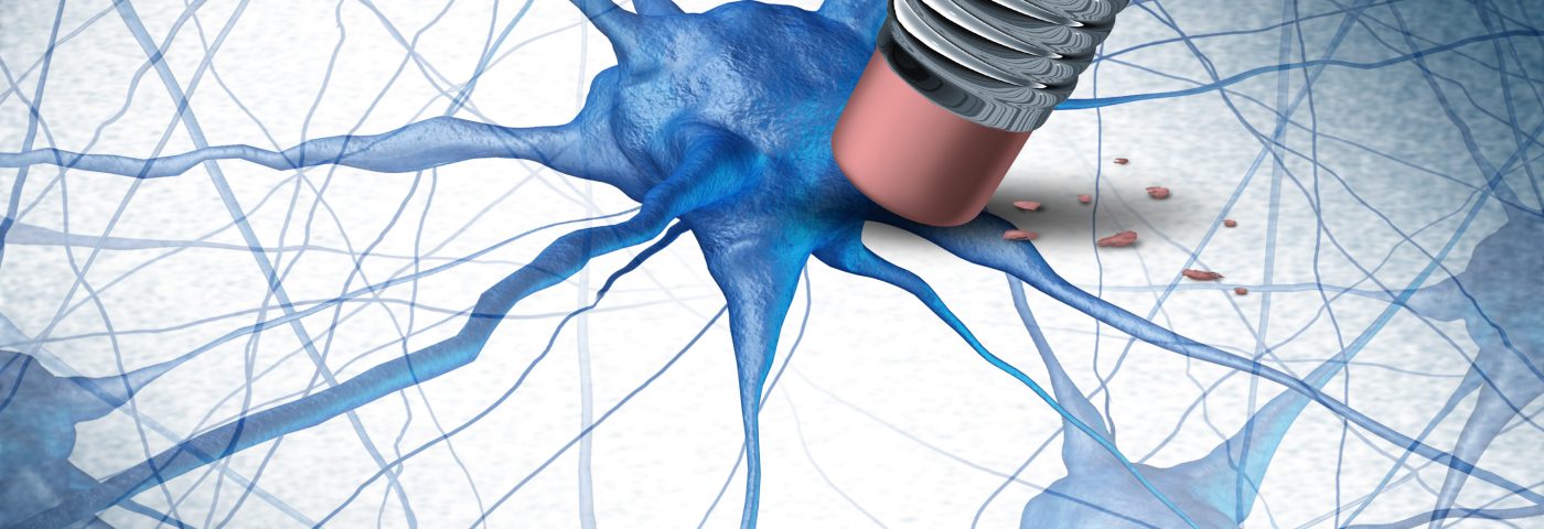
Stress granules forming inside the nerve cells of people with multiple sclerosis (MS) may be the underlying cause of nerve cell degeneration and permanent disability in these patients, researchers report.
The reason for this stress response seems to a protein that behaves aberrantly in the neurons of an MS patient, in ways that appear to trigger the formation of stress aggregates in response to inflammation.
The study, “Dysfunctional RNA binding proteins and stress granules in multiple sclerosis,” was published in the Journal of Neuroimmunology.
Hannah Salapa a PhD student at the University of Saskatchewan, Canada, and her supervisor, neurology professor Michael Levin were encouraged by the high rates of MS in Canada, and in their home province, to study what is causing, at the molecular level, nerve cell loss and consequent disability in people with this disease.
Salapa also had personal reasons, as both her grandparents were “affected by incapacitating neurological diseases,” she said in a university news article written by Federica Giannelli.
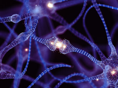
Analyzing a nerve cell line growing in the lab, the researchers found that a protein called heterogeneous nuclear ribonuclear protein A1 (hnRNP A1) stops working as it should inside nerve cells exposed to an inflammatory environment.
Antibodies against A1 and an inflammatory messenger, or cytokine, called interferon gamma (IFN-?), caused A1 to be misplaced inside nerve cells and to act abnormally once there.
A1 is vital for maintaining brain cell health. But when it no longer working properly, the researchers noted, the protein leads to the formation of stress granules (abnormal aggregates of proteins and genetic messengers called RNAs) inside nerve cells.
Importantly, the researchers were able to find these granules in brain neurons taken from a patient who, upon his death, had donated his body to science. This was the first time such structures were observed in a person with MS.
Discuss the latest research in the MS News Today forums!
“Neurons in the brain of a[n] MS patient showed pathogenic RBP [RNA binding protein] dysfunction, including nuclear depletion of hnRNP A1, its mislocalization to the cytoplasm, and its colocalization in SGs [stress granules],” the team wrote. “These data indicate a role for dysfunctional hnRNP A1 in the pathogenesis of MS.”
Results gathered suggest that the stress granules may contribute significantly to the nerve cell death and damage in the brain and spinal cord seen in MS patients and be an underlying cause of disability. Similar stress granules have also been associated with other neurodegenerative diseases, like amyotrophic lateral sclerosis (ALS), and forms of dementia.
“Knowing how the A1 protein dysfunction works may help us find treatments to reverse the effects of the A1 antibodies and inflammatory cytokines, so that we could reduce the disability caused by multiple sclerosis,” said Levin, who is also research chair of Saskatchewan Multiple Sclerosis Clinical Research.
Now, the team is planning to further study the molecular pathways that lead to nerve cell damage “and study what is the difference between healthy brains and MS brains so that we can prevent the disruption from happening,” Salapa concluded.


Karen Smith
How do you make arrangements to donate your body/brain after death occurs.