
#ACTRIMS2019 – Two MRI Biomarkers Can Potentially Distinguish RIS from Other Disorders
Written by |
Two new magnetic resonance imaging (MRI) biomarkers — called central vein sign and paramagnetic rim sign — could be useful for differentiating true radiologically isolated syndrome (RIS) patients from those with mimicking features, new research shows.
The findings were presented at the Americas Committee for Treatment and Research in Multiple Sclerosis (ACTRIMS) Forum 2019 as part of the NAIMS Symposium – The Central Vein Sign in Multiple Sclerosis: Update and Future Directions.
The March 1 presentation, titled “The Central Vein Sign and Paramagnetic Rim Sign in Radiologically Isolated Syndrome,” was given by Suradech Suthiphosuwan, MD, from St. Michael’s Hospital, University of Toronto, Canada.
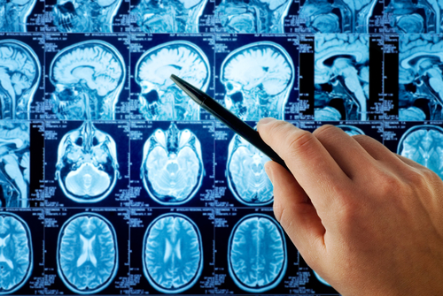
The area surrounding the brain’s central vessels — called perivenular space — is thought to be a site where an inflammatory cascade is likely triggered, leading to the formation of lesions around these veins that can be readily observed using MRI.
Although highly suggestive of MS, RIS is a disorder describing asymptomatic individuals with incidental radiologic abnormalities detected by MRI. These abnormalities can present in the perivenular space, as in MS.
According to Suthiphosuwan, “one-third of RIS cases develop clinically definitive MS within five years.”
Because of the high similarities in radiologic presentation, yet different health outcomes, there is a need to develop tests/markers able to distinguish RIS.
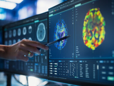
MRI studies aiming to identify MS biomarkers have demonstrated that many white matter lesions in MS patients have a visible MRI signal in the central vein — a central vein sign. These studies have also shown that some chronic MS lesions have a unique signal called the paramagnetic rim sign. This signal at the rim of lesions may represent chronic, active inflammation.
Although both central vein signs and paramagnetic rim signs have been observed in MS patients, they have yet to be assessed in RIS patients.
Thus, researchers from St. Michael’s Hospital at the University of Toronto evaluated the presence of these potential distinguishing MRI markers in white matter lesions of RIS patients.
Researchers examined 15 people with RIS (mean age of 45 years), between July 2017 and July 2018, who underwent MRI to assess white matter lesions for central vein signs and paramagnetic rim signs.
Results showed that every RIS participant had white matter lesions that were positive for the central vein sign. The average proportion of white matter lesions positive for the central vein sign per case was about four out of five lesions (83%).
In total, 14 of the 15 RIS participants had central vein signs in more than two of five white matter lesions (>40%), which meets a proposed threshold to distinguish MS from other disorders.
The paramagnetic rim sign was present in 11 out of 15 RIS participants (73%). The average proportion of white matter lesions positive for paramagnetic rim signs per individual was about two of five (19%). Paramagnetic rim signs were absent in four of the RIS participants (27%) — of these, three had low total lesion loads (9-25 white matter lesions per case), and one had the lowest proportion of positive central vein signs in white matter lesions.
A strong correlation was found between the proportion of central vein signs and paramagnetic rim signs in the white matter of RIS patients. RIS cases without paramagnetic rim signs had low proportions of positive central vein signs in white matter lesions.
Overall, most RIS cases had a large proportion of central vein sign-positive white matter lesions, indicating perivenular pathology.
“The majority of RIS cases had more than 40% central vein sign positive white matter lesions per case,” Suthiphosuwan said, adding that this result “meets the 40% threshold that has been proposed to distinguish MS from non-MS.”
“A large (though smaller) proportion of RIS cases also had paramagnetic rim sign-positive white matter lesions,” the researcher noted.
These findings suggest that the majority of RIS subjects “harbor both perivenular inflammation and subclinical chronic active demyelination similar to MS,” Suthiphosuwan added, suggesting that RIS patients “may be at risk of eventually developing progressive clinical symptoms.”
Based on the results, the team suggested that “both central vein sign and paramagnetic rim sign could potentially be useful to differentiate true RIS (i.e. subclinical inflammatory demyelination) from mimickers.”
The researchers have planned a follow-up study of this patient cohort, which will enable a better understanding of the diagnostic and predictive value of central vein and paramagnetic rim signs in RIS.

Leave a comment
Fill in the required fields to post. Your email address will not be published.