Brain Inflammation Countered by Neuronal Growth in Mouse Model of MS
Written by |

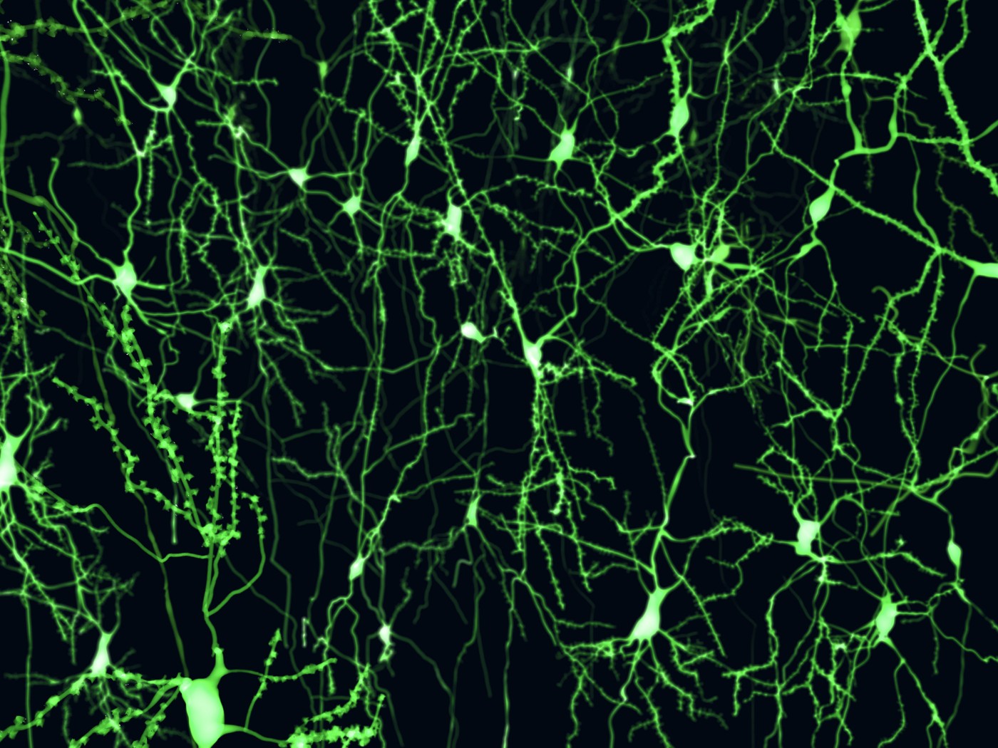
As inflammation and neuronal death progressed in the brains of mice with multiple sclerosis (MS), a molecular signaling pathway with a key player called Wnt was seen to come into action in brain areas crucial for memory production, triggering the formation of new neurons.
The findings, presented in the study “Activation of Wnt signaling promotes hippocampal neurogenesis in experimental autoimmune encephalomyelitis,” published in the journal Molecular Neurodegeneration, suggest that boosting Wnt signaling could counter the effects of brain inflammation, possibly preventing the advent of the memory and cognitive problems affecting many MS patients.
Scientists believe that as MS progresses continued neurodegeneration in the hippocampus — a brain region crucial for memory and learning — may contribute to the cognitive problems patients encounter.
Mice with experimental autoimmune encephalitis (EAE), used to model human MS processes, can tell us a lot about what is going on in the brain both during acute and chronic stages of the disease. Earlier analyses of EAE mouse brains showed that genes involved in making new neurons were activated — a somewhat counterintuitive finding since we mostly associate MS with nerve cell damage and death.
One of the genes identified is responsible for coding for Wnt (pronounced wint), a signaling molecule involved in tissue renewal and regeneration. Again, using mice with EAE, researchers at Heinrich Heine University Düsseldorf, in Germany, tracked changes in Wnt and the birth and death of neurons.
In the acute stages of EAE in the mice, researchers found that Wnt signaling increased in the hippocampus, and the increase was found particularly in areas where neurons were dying. These areas also had high levels of immune cytokines such as TNF-alpha and IFN-gamma, suggesting that inflammation could be a factor activating Wnt.
To test this idea, researchers used brain slices, which they kept alive in a lab dish, treating them with TNF-alpha. As they suspected, Wnt signaling increased in brain tissue exposed to the inflammatory factor.
Most important, however, was the finding that the increased Wnt signaling spurred the growth of new neurons, both in mice and in cells grown in the lab. If cells were sprinkled with TNF-alpha while blocking Wnt, no new cells were born.
Researchers also found that the process included not only nerve cells, but also microglia and astrocytes, glial cells that are essential to brain function.
Future experiments will reveal if boosting Wnt signaling might be used as a therapy to promote nerve cell growth in areas affected by brain inflammation.


