
Novel MRI Marker Better at Predicting MS Progression, Study Reports
Written by |
A large retrospective study suggests that a magnetic resonance imaging (MRI) marker — called “brain atrophied T2 lesion volume” — could help predict the timing of multiple sclerosis (MS) progression.
According to the study, this marker was the only MRI parameter capable of predicting disease progression, compared with other markers such as changes in number and volume of lesions, or whole brain atrophy (shrinkage).
The study, “Atrophied Brain T2 Lesion Volume at MRI Is Associated with Disability Progression and Conversion to Secondary Progressive Multiple Sclerosis,” was published in the journal Radiology.
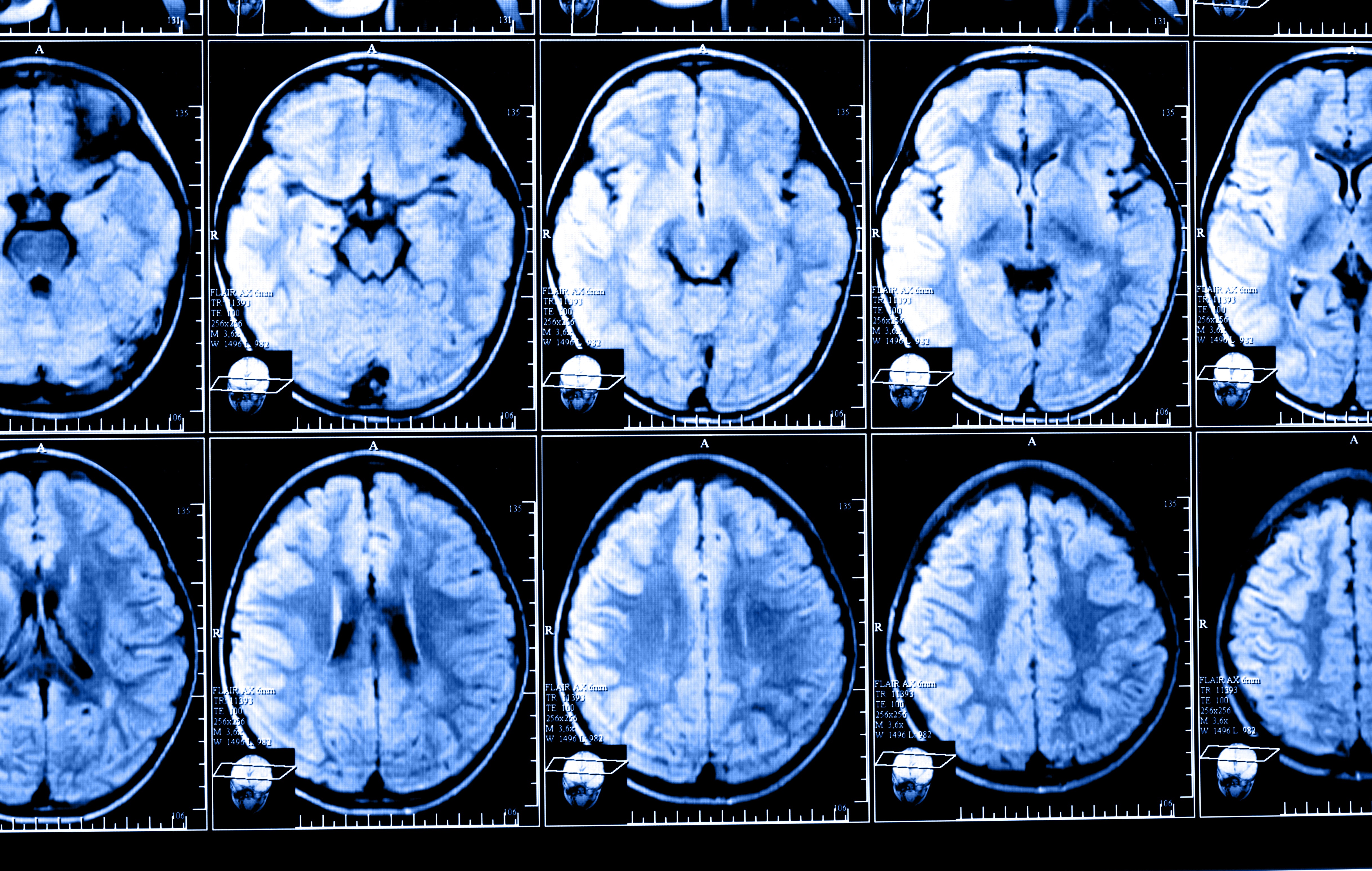
MRI imaging is routinely used for surveillance of abnormal changes in MS patients. By quantifying the number and volume of new lesions, clinicians are able to estimate active inflammation in a quantitative way.
In clinical trials testing new therapies for MS, the U.S. Food and Drug Administration commonly asks researchers to assess the ability of a new treatment to reduce the number of lesions over 24 months. However, the association between MRI markers and worsening of MS disease has not been well established.
Atrophied T2 lesion volume is a measure of the number of lesions being replaced with cerebrospinal fluid spaces as a result of atrophy (brain shrinking) or direct lesion destruction.
To study whether this marker is in fact a good indicator of MS progression, researchers at the Jacobs School of Medicine and Biomedical Sciences, at the University of Buffalo, collected data on a large group of patients during a five-year follow-up period.
Overall, researchers analyzed MRI scans from 1,314 MS patients (mean age of 46 years), 124 patients with clinically isolated syndrome (CIS; mean age of 39 years), and 147 healthy controls (mean age of 42 years).
Results showed that 336 patients with CIS or MS showed disease progression, and 67 converted from CIS or relapsing-remitting MS (RRMS) to secondary progressive MS (SPMS).
Patients with disease progression accumulated a greater amount of atrophied T2 lesion volume per year during the study. When researchers compared this MRI marker between patients with or without disease progression, they observed that patients with disease progression had on average 34.4 cubic millimeters more of atrophied T2 lesion volume, than those whose disease didn’t progress.
The researchers estimated that an increase of 1 mL (1000 cubic millimeters) in the annualized atrophied T2 lesion volume was associated with a five times higher risk of disease progression. Additionally, for every 1 mL increase in this marker, there was a 22.8% higher chance for a disease progression event.
Similarly, patients who progressed to SPMS also had greater annualized atrophied T2 lesion volume — a difference of more 26.4 cubic millimeters on average. For these cases, the researchers also calculated that an increase of 1 mL in atrophied T2 lesion volume was associated with a 4.7 times higher risk for progressing to SPMS.
Of note, the team found that differences in the percentage of brain volume change, percentage of ventricular volume change, or number of lesions were not able to predict the conversion to SPMS.
“Neither changes in number and volume of lesions nor the development of whole brain or central brain atrophy showed any predictive power in demonstrating which patients would progress to secondary progressive MS, either from initial presentation of the disease, called clinically isolated syndrome, or the next stage, relapsing-remitting MS,” Robert Zivadinov, MD, PhD, director of the Buffalo Neuroimaging Analysis Center, and study’s leading author, said in a press release.
“The fact that atrophied lesion volume was the only measure that was predictive of conversion to progressive multiple sclerosis, and brain atrophy was not, is a major novel finding of this study,” he added.
This new finding is in line with a previous study in which Zivadinov’s team showed that atrophied brain lesion volume was a better parameter for assessing disease progression, in comparison with the appearance of lesions or whole brain atrophy.
“This study corroborates initial reports from our group regarding using atrophied lesion volume as a potential MRI marker of disease progression in a large, population-based cohort of MS patients followed in clinical routine,” said Zivadinov.
Overall, the team said, “in this large population-based cohort study, we showed that atrophied brain T2 lesion volume represents a viable predictive MRI marker of the development of disability progression and conversion to secondary progressive multiple sclerosis disease course.”


Jeff
Interesting information. It may be a typo but milliLITERS are not comparable to milliMETERS.
Milliliters measure volume, millimeters measure length. Cubic Millimeters are not comparable to cubic milliliters.