
New Imaging Agent of Myelin for Earlier MS Diagnosis, Myeliviz, Entering Clinical Testing
Written by |
The U.S. Food and Drug Administration (FDA) has agreed to allow Myeliviz, an imaging agent of myelin — the protective layer that covers nerve fibers and is damaged in multiple sclerosis (MS) — to be evaluated in a clinical trial with healthy volunteers.
Myeliviz, created by Case Western Reserve University researchers, has the potential to be used in diagnosing MS, as well as other myelin-associated diseases, and in monitoring disease progression.
“Myelin has never been directly imaged before. Our technique is the first to do so, and we are hopeful that this will provide earlier and more accurate diagnosis of MS,” Yanming Wang, PhD, Myeliviz’s co-inventor and a professor of radiology at the Case Western Reserve School of Medicine, said in a press release.
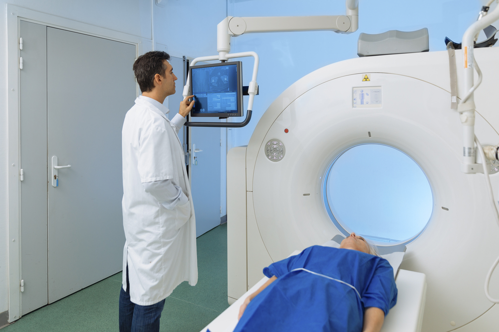
Myeliviz is injected intravenously (directly into the bloodstream) into a patient, and travels through the blood to target and bind to myelin, “lighting it up” during positron emission tomography (PET) imaging scans. Areas of low or absent “light” on these PET scans would correspond to areas of myelin loss.
Preclinical studies showed that Myeliviz can directly detect and quantify myelin changes in the brain and spinal cord of rodents with good spatial and temporal resolution, according to a previous press release by the Cleveland Clinic, which collaborated with Case Western in developing the agent.
Researchers believe that PET scans with Myeliviz may supplement, and in some cases replace, magnetic resonance imaging (MRI) scans, the current gold standard imaging technique for MS. While MRI scans do not assess myelin directly, they allow experts to detect brain lesions associated with the loss of myelin.
MRI scans are currently the most sensitive, non-invasive tool for diagnosing MS. However, they are less effective in monitoring disease progression.
Robert Fox, vice chair for research at the Cleveland Clinic’s Neurological Institute, said “the goal is to enable clinicians to more unambiguously diagnose MS and monitor disease progression and repair processes.”
Myeliviz’s potential will now be evaluated in volunteers at the Cleveland Clinic Mellen Center for Multiple Sclerosis, making it the first myelin-PET imaging agent to be studied in people. This study will assess the agent’s safety and pharmacokinetics in the body (its absorption, distribution, metabolism and excretion), and provide evidence of early efficacy by comparing Myeliviz PET measures to those of conventional MRI.
The researchers anticipate that positive results from this trial will support a Phase 2 study in MS patients.
The clinical program of Myeliviz is being supported by a $1.6 million grant from the National Institutes of Health, with Wang and Fox as the grant’s co-principal investigators.
Researchers believe that Myeliviz could help in the development of new MS therapies as a measure of treatment effectiveness, and may significantly aid in slowing disease progression, as treatment could be started earlier. Early stages of MS are more responsive to neuroprotective therapies.
“Myeliviz could be the missing link in finding a cure for MS and other myelin diseases by serving as a specific and quantitative imaging marker for early diagnosis and sensitive, quantitative evaluation of novel therapies currently under development,” said Chunying Wu, Myeliviz’s co-inventor and an instructor of radiology at Case Western.
Mykol Larvie, the Cleveland Clinic’s director of functional neuroimaging, added that Myeliviz could be used to monitor “brain health in general,” and to help evaluate the effectiveness of treatments in “numerous neurological diseases such as epilepsy, stroke, neurodegeneration, tumor and trauma in the brain and spinal cord.”


Leave a comment
Fill in the required fields to post. Your email address will not be published.