New Nanosensor May Help to Diagnose MS at Early Stages
Written by |

A new tiny sensor is able to detect antibodies against myelin, the protective coating of nerve cell axons whose destruction is a hallmark of multiple sclerosis (MS), potentially allowing for a diagnosis in early disease stages, researchers report.
It also offers the possibility of distinguishing multiple sclerosis from neuromyelitis optica, a rare autoimmune inflammatory condition that shares symptoms with MS.
Their study, “Nanoimmunosensor based on atomic force spectroscopy to detect anti-myelin basic protein related to early-stage multiple sclerosis,” was published in the journal Ultramicroscopy.
The immune system of MS patients recognizes myelin as foreign, launching damaging attacks on myelin in the central nervous system (brain and spinal cord) and disrupting nerve cell communications via the transmission of electrical impulses.
Demyelinating diseases like MS are hard to confirm, as the diagnosis often relies on reported symptoms and MRI scans to detect brain lesions.
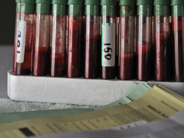
A team led by researchers at the Federal University of São Carlos (UFSCar), in Brazil, developed a nanosensor that is able to detect myelin-targeting antibodies in small amounts of cerebrospinal fluid, or CSF. This fluid surrounds the brain and spinal cord, and is usually collected through a spinal tap.
To develop this sensor, the researchers adapted a technology called an atomic force microscopy (AFM; also called atomic force spectroscopy ) that was originally used to detect herbicides, heavy metals, and other toxic compounds. AFM is a very sensitive technique that is able to measure interaction between molecules.
AFM was used to test whether samples from MS patients contain antibodies against myelin basic protein (MBP) — the key protein in myelin — and if their presence could be a biomarker of MS.
In this application, MBP peptides (short proteins) are attached to a chemically modified surface, and used as a substrate where antibodies from patient samples can be immobilized. The peptides, called MBP 85-99, used in the study were synthesized by a laboratory at the University of São Paulo.
“Atomic force spectroscopy can detect the presence of specific antibodies for … [MS and neuromyelitis optica] in cerebrospinal fluid and blood serum. If the antibodies are attracted by the peptides deposited on the sensor during the test, this is a sign that the patient has the disease,” Fabio de Lima Leite, a researcher at the Science and Technology for Sustainability Center at UFSCar, and the study’s lead author, said in a press release.
“The device is highly sensitive and can detect a small amount of antibodies, so the method can diagnose the disease at an early stage,” he added.
Researchers analyzed samples from five patients with relapsing-remitting MS (RRMS) at different disease stages and five people without demyelinating conditions, all being followed at the hospital of São Paulo State University’s Botucatu Medical School.
Before AFM measures were taken, samples were purified to isolated patients’ antibodies.
“The cerebrospinal fluid and serum were purified, leaving only antibodies in each sample. This enabled us to detect specific antibodies for multiple sclerosis, such as anti-MBP 85-99. If these antibodies are circulating in a patient, they probably have multiple sclerosis,” Lima Leite said.
Results showed that the sensor had a 96.68% sensitivity, and a 68.48% specificity to detect MS. A test’s sensitivity refers to its ability to correctly identify those with a given disease, while specificity refers to correctly identifying those without it.
The sensor’s sensitivity is superior to previous techniques (like ELISA) that detected antibodies against MBP peptides with a sensitivity of 83.7%. It was also able to clearly distinguish MS patients at early stages from those with more advance disease.
“There’s no cure, but early diagnosis can give patients quality of life and better treatment,” said Ariana de Souza Moraes, a UFSCar researcher and study author.
Lima Leite also noted that the new technique is less costly for clinics to acquire than conventional MRIs. “Our method is more accurate, avoiding diagnostic errors, as well as being cheaper. An AFM [atomic force microscope] can cost about 20,000 dollars, whereas an MRI machine costs upwards of 400,000 dollars.”
Furthermore, the sensor can easily be adapted to accurately diagnose neuromyelitis optica and exclude MS.
In a previous study, researchers identified aquaporin 4 as a diagnostic biomarker for neuromyelitis optica. “A biomarker for the disease exists, so it was possible to detect the anti-aquaporin 4 antibody in patient samples by the same method as that used to detect multiple sclerosis,” Moraes said.
“The two disorders have similar symptoms but different action mechanisms and treatments,” Moraes added. “An incorrect diagnosis can aggravate the disease. If a patient with neuromyelitis optica is treated for multiple sclerosis, optic nerve inflammation is accelerated and can’t be reversed. The sensor is expected to represent a major advance for patients with demyelinating disorders.”
Researchers now aim to develop a sensor that works without the need to purify patient samples.