
New wearable microscope can help image cell activity in mice
Worn by the mice, this tool could prove valuable in MS research
Written by |
A team of scientists has developed a wearable microscope — to be carried on the backs of animals in a lab — that can be used to evaluate cell activity in the spinal cord of living mice with greater clarity than has ever before been possible.
The scientists say this new technology could be a valuable research tool for studying neurological diseases such as multiple sclerosis (MS), which is caused by inflammation that damages the central nervous system, comprised of the brain and spinal cord.
“Our wearable microscopes fundamentally change what is possible when studying the central nervous system,” Axel Nimmerjahn, PhD, a professor at Salk Institute, in California, said in a press release.
Pavel Shekhtmeyster, PhD, a former postdoctoral fellow in Nimmerjahn’s lab and co-first author on the new studies, noted that the team’s researchers have “overcome field-of-view and depth barriers in the context of spinal cord research.”
“Our wearable microscopes are light enough to be carried by mice and allow measurements previously thought impossible,” Shekhtmeyster said.

Wearable microscope may ‘revolutionize the study of pain’ in MS
The spinal cord plays a vital role in relaying nerve signals between the brain and the rest of the body. Signaling in the spinal cord is thought to involve a complex ballet of interconnected cellular processes — but technological limitations have made it difficult to study these processes in living animals during laboratory research.
The design of the team’s new wearable microscope was detailed in the study, “Trans-segmental imaging in the spinal cord of behaving mice,” published in Nature Biotechnology.
The scientists built on prior advances in technology to design a microscope that could image cells in live mice with a field of view around 3.2 by 2.5 millimeters. That field is wide enough to be able to image multiple segments of the mouse spinal cord at the same time — something that has never been possible with previous technology.
“These new wearable microscopes allow us to see nerve activity related to sensations and movement in regions and at speeds inaccessible by other high-resolution technology,” Nimmerjahn said.
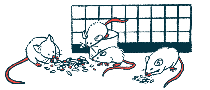
The microscopes weighed slightly less than 10 grams — less than half an ounce. They were light enough that even relatively young mice could carry them without difficulty.
“Remarkably, 8- to 16-week-old mice could carry the device on their backs without extensive training,” the researchers wrote, noting it took 30 minutes of habituation.
The microscopes had a resolution of less than three micrometers, which is more than detailed enough to be able to view individual cells with reasonable clarity. The researchers noted that the resolution of the microscope isn’t quite able to see smaller-than-cell structures, like synapses, which are the individual connections between nerve cells. Thus, additional work could further improve the device.
Our wearable microscopes are light enough to be carried by mice and allow measurements previously thought impossible.
To illustrate the utility of the system, the researchers used it to image mice that had been genetically engineered so that certain cells, called astrocytes, in their bodies would give off a fluorescent signal when active.
Astrocytes are star-shaped cells in the central nervous system that are thought to be involved in coordinating a range of neurological activities. However, these cells often go awry in MS, contributing to the inflammation and nerve cell damage that marks the condition.
The researchers demonstrated that when the mice were given a painful stimulus — a pinch on the tail — there was widespread astrocyte activation in the spinal cord. This activation did not occur when the mice were running around without pain.
The finding suggests that astrocyte activation plays a key role in driving the sensation of pain. Better understanding these processes may open avenues for the treatment of pain in MS and other neurological disorders, the researchers said.
“Being able to visualize when and where pain signals occur and what cells participate in this process allows us to test and design therapeutic interventions,” said Daniela Duarte, a study co-author at Salk.
“These new microscopes could revolutionize the study of pain,” Duarte said.

Using the wearable microscope to get a deeper view
While the microscope itself is a step forward, a limitation of using this system alone is that it can only view cells at the top of the mice’s spinal cord. In a separate study, the researchers illustrated how a similar system could be used to view cells deeper in the spinal cord.
That study, “Multiplex translaminar imaging in the spinal cord of behaving mice,” was published in Nature Communications.
This system involves the use of small reflective objects called microprisms, which are surgically implanted into the animal’s spine. Conceptually, the system works a bit like how a mirror can be used to look around corners — the microscope uses the reflective microprism to image deeper structures that the microscope itself can view.
“The microprism increases the depth of imaging, so previously unreachable cells can be viewed for the first time,” said Erin Carey, a researcher at Salk and a study co-author.
“It also allows cells at various depths to be imaged simultaneously and with minimal tissue disturbance,” Carey said.
Using this system, the researchers found that pinching the mice’s tails tended to lead to activation of astrocytes in one specific area of a part of the spinal cord: the dorsal horn. By contrast, when mice were running around, astrocytes in a different area of the dorsal horn tended to become active.
“Our data show that acute pain and animal locomotor activity evoke astrocyte [activation] in distinct dorsal horn regions,” the researchers concluded, noting that further assessment to understand these mechanisms could help identify therapies for pain and other neurological problems.
The team also noted that repeated imaging through the microprisms was feasible for at least four weeks, which “may enable the study of prolonged structural and functional biological processes,” such as MS progression, nerve pain, and responses to treatments.
The work was supported in part by the National Institutes of Health.
Leave a comment
Fill in the required fields to post. Your email address will not be published.