
Myelin may promote nerve cell damage in early immune attacks
More 'irreversible damage' with inflammation seen with fuller myelin sheath
Written by |
Nerve cells coated with myelin — the fatty substance that’s lost in multiple sclerosis (MS) — may be more vulnerable to degeneration in an inflammatory environment than cells lacking myelin, researchers working in MS patient tissues and mouse models report.
The scientists believe the phenomenon arises when certain cells responsible for supporting myelinated nerve projections, or axons, become dysregulated in the early autoimmune environment.
Their findings are in contrast to the long-standing belief that myelin is always protective, and its loss in MS is a “principal cause” of axonal degeneration.
“By challenging the prevailing image of myelin as a solely protective structure, we can gain a deeper understanding of the disease and potentially develop new treatment strategies that will maintain the functionality of the axons,” Ruth Stassart, MD, a professor at the Leipzig University Hospital in Germany and a study author, said in a university press release.

Axon damage evident at early MS stages, before substantial loss of myelin sheath
The study, “Myelin insulation as a risk factor for axonal degeneration in autoimmune demyelinating disease,” was published in Nature Neuroscience.
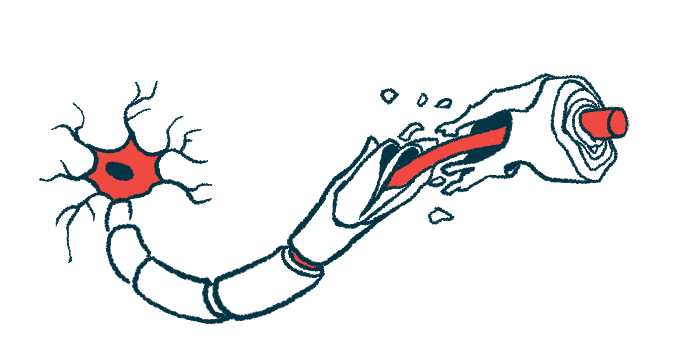
Myelin is a fatty substance that surrounds axons, the long fiber-like projections helping neurons to send electrical signals faster. It is generally considered to be a protective structure, preventing nerve cell damage and ensuring quick cell-to-cell communication.
The fatty coating is produced by a set of support cells called oligodendrocytes, which also provide myelinated axons with metabolic and structural support, including nutrients for their energetic needs and antioxidants to combat cellular stress.
An autoimmune disease, MS is characterized by the immune system’s mistaken attack on myelin. It has long been considered that myelin loss, or demyelination, leaves axons vulnerable to local inflammatory processes and is the main cause of the disease’s irreversible nerve cell degeneration.
Still, axon damage can be observed in very early disease stages — even before substantial demyelination occurs — suggesting that other factors might contribute to axonal vulnerability in MS, according to the scientists.
Researchers at Leipzig University and the Max Planck Institute for Multidisciplinary Sciences in Göttingen wondered whether inflammatory activity early in MS might disrupt the ability of oligodendrocytes to properly support myelinated nerve cells, thus contributing to axon degeneration.
Should that be true, myelinated cells — which are dependent on oligodendrocytes for support — would be more vulnerable to degeneration than cells that don’t have myelin.
Inflammation may affect ability of oligodendrocytes to support axons
“We reasoned that autoimmune inflammation may disrupt oligodendroglial support mechanisms and hence primarily affect axons insulated by myelin,” the researchers wrote.
To test this, they examined axon damage more closely in patients’ biopsy tissues and mouse MS models.
In human MS lesions, the scientists identified two types of axon damage — one which was likely reversible, and another corresponding to irreversible damage. Notably, while reversible damage was common in both myelinated and demyelinated axons, damage beyond repair was found exclusively in axons still coated with myelin.
Additional experiments in mouse MS models were consistent with tissue findings. The scientists again found these two types of axon damage in areas of inflammation, with myelinated axons more susceptible to irreversible damage.
Efficient demyelination of axons in response to autoimmune activity also resolved reversible damage to nerve cells before it progressed beyond the point of recovery.
Moreover, mice in an MS model genetically engineered to have more unmyelinated axons than normal had a less severe clinical course.
While normally protective, insulation from myelin in the early inflammatory MS environment seems to raise the risk of axon degeneration. Indeed, several alterations to oligodendrocytes can be observed in mouse MS models, indicating they may have a lower capacity to support myelinated nerve cells, the researchers wrote.
Considering these findings, the scientists proposed that “the normally symbiotic relationship between the axon and the myelinating oligodendrocyte turns into a fatal one upon disease onset.”
Work needed into mechanisms allowing injured oligodendrocytes to do damage
“Untangling the molecular mechanisms by which immune-injured myelinating oligodendrocytes convey axonal damage may be key to identifying new axo-protective therapeutic targets in multiple sclerosis,” they added.
The scientists noted that while demyelination in the short term might help to avoid axon damage, chronic demyelination likely comes with other consequences. The work of healthy oligodendrocytes that remyelinate cells after inflammation has resolved is essential for the health of nerve cells, they said.
“Taken together, our study suggests that not demyelination per se, but oligodendroglial integrity and downstream axonal support may constitute crucial future therapeutic targets for acute autoimmune demyelinating diseases such as multiple sclerosis,” the researchers concluded.
“Instead of preserving the damaged myelin, it might actually be therapeutically better to promote rapid degradation of damaged myelin and support the regeneration of functional myelin,” added Robert Fledrich, a researcher in the Institute of Anatomy at Leipzig University.


Leave a comment
Fill in the required fields to post. Your email address will not be published.