
Synapses in CNS may be important in myelin production, per MS study
Better understanding their role may lead to new disease treatment strategies
Written by |

Junctions between nerve cells and certain cells called oligodendrocyte precursor cells, or OPCs, in the brain and spinal cord may play an important role in producing myelin, the protective substance that’s progressively lost in multiple sclerosis (MS), new research suggests.
OPCs are abundant in the brain and spinal cord and play various roles in brain health. A subset of them mature into oligodendrocytes, the cells responsible for producing myelin in the central nervous system (CNS), comprised of the brain and spinal cord.
Insights into the function of these poorly understood connections, or synapses, could eventually be leveraged to develop therapeutic strategies for people with MS and other brain diseases, according to the scientists.
“This [research] gives an understanding of the basic, fundamental properties of how these cells work in normal development. In the future, we might look at how they function differently in the context of MS patients,” Kelly Monk, PhD, professor at the Oregon Health & Science University (OHSU) and the study’s senior author, said in a university press release.
The findings were detailed in a study, “Synaptic input and Ca2+ activity in zebrafish oligodendrocyte precursor cells contribute to myelin sheath formation,” published in Nature Neuroscience.

Investigating the role of OPC-nerve cell connections
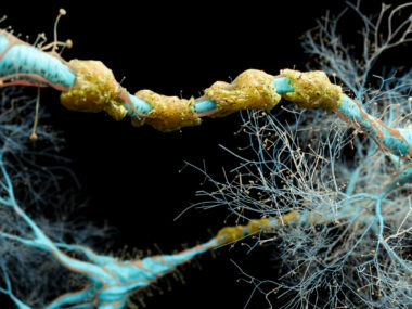
The myelin sheath is a fatty substance that surrounds nerve cell projections, protecting them and helping to speed the transmission of electrical signals to other cells. In MS, self-reactive immune attacks cause the destruction of myelin, leading to impaired nerve communication and nerve damage.
Research suggests that dysfunction of these OPCs could contribute to MS disease processes. Understanding their role and relationship to neurons, therefore, could be important for developing therapeutic strategies for MS.
More than 20 years ago, OHSU researchers made the discovery that OPCs form synapses with neurons. Synapses are the site where two nearby cells directly communicate with one another via chemical signals.
Since then, however, little has been uncovered about the function of these synapses. No study had ever before looked at their activity in a live animal model.
“After two decades, we still didn’t know what these synapses do,” Monk noted.
To learn more, the researchers now aimed to look more closely at the real-time function of OPC-neuron synapses in the spinal cord of zebrafish, using single-cell imaging and a range of other experimental techniques. With many nervous system features similar to those seen in humans, zebrafish are commonly used in neurodegenerative disease research.
OPC synapses were found to be composed of puncta, or clusters, of certain proteins, namely MAGUK/PSD-95 and gephyrin. These puncta were found close to markers normally seen in the nerve cell side of a synapse, supporting the presence of a connection between the two cell types.
The formation of gephyrin puncta was dynamic — assembling and disassembling often over time — but typically repeatedly formed in the same locations, which the researchers dubbed “hotspots.”
Importantly, these hotspots were found in the proximity of areas where myelin would later form. About 30% of the hotspots in OPCs predicted a future myelin sheath location in that area.
We propose that neuron-OPC synapses are dynamically assembled and can predetermine myelination patterns.
This relationship appeared dependent on chemical communication from the neuron side of the synapse. When that communication was inhibited, or blocked, OPC hotspots were no longer able to predict future myelination.
Calcium signaling activity in the OPCs also was found to predict future myelination, which was similarly regulated by signals from neurons.
Genetic disruption of various proteins important for OPC synapse function led to the development of fewer oligodendrocytes and a reduction in the myelin sheath.
Altogether, the findings suggest that neuronal activity influences OPC activity to regulate future myelination patterns.
“We propose that neuron–OPC synapses are dynamically assembled and can predetermine myelination patterns,” the researchers wrote.

Altering OPC function in MS might be way to promote myelin restoration
More work will be needed to understand which chemical signals from neurons are important for this relationship, and to know the cellular events downstream of OPC calcium signaling that influence myelin formation.
According to Jiaxing Li, PhD, a postdoctoral fellow in Monk’s lab and the study’s first author, these new discoveries likely mark only the first steps in understanding the role synapses play in myelin production.
But scientists may be able to use the findings to develop new therapeutic strategies for various diseases, including MS, per the team.
Indeed, according to the researchers, altering OPC function in MS could be a way of promoting myelin restoration, or remyelination, and thereby slowing disease progression.
More broadly, the scientists believe the findings highlight the likely importance of OPC-neuron connections in the brain beyond myelin formation. One point for further research is that, while OPCs account for about 5% of all brain cells, only some of them go on to become oligodendrocytes.
“It’s becoming pretty clear that these OPCs have other functions aside from forming oligodendrocytes,” Monk said, adding that, “from an evolutionary perspective, it doesn’t make sense to have so many of these precursor cells in your brain if they’re not doing something.”
The scientists believe future research should also explore these possible other roles for the OPC-neuron synapse.
This work was supported by a National Multiple Sclerosis Society postdoctoral fellowship, a Warren Alpert Distinguished Scholar Award, and awards from the National Institute of Neurological Disorders and Stroke of the National Institutes of Health. All research involving animals was cleared by OHSU’s Institutional Animal Care and Use Committee.

Leave a comment
Fill in the required fields to post. Your email address will not be published.