
Professor Earns Research Award for Establishing Use of MRI to Improve MS Diagnosis, Understanding
Written by |
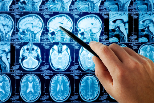
Professor Frederik Barkhof, MD, PhD, has won the 2018 John Dystel Prize for Multiple Sclerosis Research for pioneering the use of magnetic resonance imaging (MRI) to improve multiple sclerosis (MS) diagnosis and understanding of the disease.
The prize, decided by a peer committee, is awarded annually by the National MS Society and the American Academy of Neurology (AAN). It was created in 1994 in honor of John Jay Dystel, who died from complications related to MS.
Barkhof will receive the award at a ceremony April 22 at the 2018 AAN Annual Meeting in Los Angeles, where he will also give a 20-minute lecture. The prize also includes $15,000 to be used at the winner’s discretion, and complimentary registration and expense reimbursement for the meeting.
“In my opinion, no advance in MS research has made more of an impact on the disease than MRI and Professor Barkhof has been at the forefront of this effort since the early stages of its development,” Henry F. McFarland, MD, the 1998 Dystel Prize winner, said in a press release.
Barkhof is a professor of neuroradiology at VU University Medical Center in Amsterdam and at University College London in London.
Among his contributions is the development of MRI criteria for MS diagnosis. His team defined guidelines to focus on specific brain regions to look for MS-related lesions, which are known as the Barkhof criteria. These guidelines enable an earlier and more reliable diagnosis, and are the basis of today’s MS diagnostic strategy.
He also contributed to the expanding use of imaging-based clinical trials, which allow for better analysis of nerve protection and repair, disease progression and disability, and effectiveness of treatment candidates.
Barkhof was also a leader in the use of measures for T1-“black-hole” hypointense brain lesions. These areas of relatively severe damage, considered markers of the disease process, show up differently on MRIs from the more nonspecific T2 hyperintense lesions.
“His insights have greatly contributed to modern trial design and therapeutics, which is now propelling the field in important and increasingly focused directions,” said Jerry S. Wolinsky, MD, an Emeritus Professor in Neurology at McGovern Medical School, University of Texas.
Barkhof has also authored three books: “Magnetic Resonance in Dementia,” “Clinical Applications of Functional Brain MRI,” and “Neuroimaging in Dementia” as well as chapters in several books. He’s also written more than 900 peer-reviewed articles, and was named one of the 3,000 most influential scientists worldwide by Thomson Reuters.
As an early leader of the MAGNIMS consortium, a European network of academics who use MRI to study MS, Barkhoff helped establish and define the role of MRI in MS diagnosis and research. He has also given several lectures on MS around the world, and mentored numerous students.
“He has made important contributions across a range of interrelated areas including diagnosis, monitoring, trial design, and disease mechanisms,” said Alan J. Thompson, MD, the 2017 Dystel Prize winner. He combines “collegiality with scientific rigor and a passion for collaboration.”


Violet Weinberg
It is interesting there is no comment on the difference between distinguishing new lesion activity on Tesla 3 versus Tesla 1.5, etc. MRIs. For instance the new drug Ocrevus is not approved for use for patients diagnosed with Secondary Progressive MS - because there is no evidence of disease progression as seen on Tesla 1.5. However MS patients, who have had the resources to pay for the advanced Tesla 3 have found new lesions, unseen on the 1.5. They, then would or should be approved for Ocrevus as their diagnosis of SPMS is not accurate. Thus a number of physicians, (neurologists) have listed their SP patients on the Ocrevus applications as having "Primary MS" in order to get treatment. Unfortunately since their lesions do not show evidence of increased disease before they begin the program, they also do not show decrease after the program (as checked after a year of treatment)........so there this is a "hope this may have helped even though we had no way of assessing need in the first place, nor any changes after treatment..........
Carol Moore
Please consider Dr. Raymond Damadian and Dr. Scott Rosa for next year’s award. They are using FONAR’s Upright MRI and the Image Guided Atlas Treatment to cure Multiple Sclerosis. I am one of their patients and I was able to stop taking Rebif in 2012, have not had a flare since and have more energy, stamina and neurological function than I did as a kid. I am 41 years old now. These doctors gave me back my life without surgery or drugs.
Deborah Tefertiller
Wow Carol! I am newly diagnosed and going to start on Tecfidera when funding is approved. I am 57 and VERY much interested in hearing more about this treatment regimen! Can you tell me where to read more about it? I have never heard of it in all the research I have been doing.