
#EAN2018 – Pupil Response to Light Linked to RRMS Duration and Severity in Study
Written by |
Measuring the response of the pupil to light stimulating the eye is a non-invasive and easy way to assess multiple sclerosis (MS) severity and progression, researchers report.
A clinical study found that poor, or dysfunctional, pupil response was associated with longer disease duration and greater disease severity in relapsing-remitting multiple sclerosis (RRMS) patients.
These findings were presented at the 4th Congress of the European Academy of Neurology (EAN) that recently took place in Lisbon. The presentation was titled “Reduced pupillary modulation in patients with relapsing-remitting Multiple Sclerosis is associated with longer disease duration and higher disease severity” (abstract on page 80).
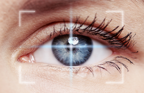
The pupil, the black spot at the center of the colored structure (iris) of the eye, controls the amount of light that enters the eye by contracting (closing) or expanding (dilating). When the eye encounters a strong light, the pupil rapidly contracts — and when little light is available, the pupil widens or expands to allow as much light as possible to enter.
Abnormal pupil reactions to light are common among RRMS patients, and believed to be due to MS lesions in the nervous system. Still, the extent to which this visual manifestation of MS correlates with disease duration and severity is not fully understood.
Researchers at the University of Erlangen-Nuremberg, in Germany, examined 85 RRMS patients (mean age 37.4), and evaluated their pupil’s diameter, dilation and constriction velocity, and reflex response.
They found that the pupil’s diameter and “early re-dilatation velocity” — speed at which the pupil reacted immediate to strong light — negatively correlated with a patient’s age, disease duration, and disability scores. This means that the worse result in these tests were seen in older RRMS patients, those with longer disease duration, and those with worse disability.
Repeatedly quick pupil response — or early re-dilation velocity — was positively associated with the functional status of the patients, meaning that better results here reflected lesser disability, in terms of both movement (walking and ambulation, hand and arm function, etc.) and cognition.
Slower and poorer pupil response to light was linked to longer disease duration and older age, as well as worse disability and functional scores.
Based on these results, the team concluded that “the decrease in sympathetic and parasympathetic pupillary autonomic modulation was associated with longer disease-duration and higher disease-severity.”
Because pupil features and reactivity can be easily and non-invasively examined using light reflex pupillography, results seen with such tests should make them an attractive clinical marker of “MS severity and progression,” the researchers suggested.


Rachelle
Hi I am 25 was diagnosed in Sept 2016 I suffered what they called optic neuritis in my left eye it took over a year to gradually get better but is not 100% I now have it in my right eye... Can you explain what your saying in this article please thanks
Alice Melão, MSc
Dear Rachelle,
Optic neuritis is an inflammation of the optic nerve and not an alteration of the pupil response.
In this study the researchers confirmed that RRMS patients had impaired pupil response, and that this alteration was associated with disease severity and functional disability. However, they did not explored a potential association between impaired pupil response and what you had, optic neuritis.
Jeff Wislon
My eyesight comes and goes is this related to MS? What can be done?
Alice Melão, MSc
Dear Jeff,
We at Multiple Sclerosis News Today are not medical doctors, and so we can not do clinical evaluations of your symptoms. You should discuss your visual alterations with you physician or go to a eye specialist.
Still, it has been recognize that MS patients may experience more frequently optic neuritis - an inflammation of the optic nerve, and alterations of the pupil response, both of which may affect visual acuity.