
Brain Iron Levels Correlate with MS Progression, Disability Risk, Study Shows
Written by |

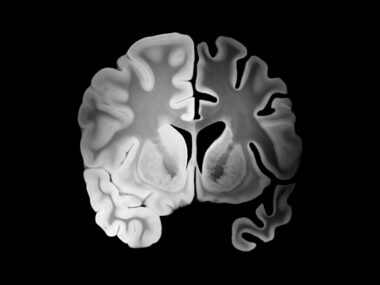
Evaluating the local differences in iron accumulation in the deep gray matter of the brain using a special magnetic resonance imaging (MRI) technique, may help identify multiple sclerosis (MS) patients at greater risk for disease progression and disability, a study reports.
The study “Brain Iron by Using Quantitative MRI Is Associated with Disability in Multiple Sclerosis” was published in the journal Radiology.
Measuring brain’s atrophy (shrinkage) is currently used as a predictor of cognitive and physical disability in MS, but assessing these changes in the brain is a long-term task.
“Brain atrophy takes a long time to see,” Robert Zivadinov, MD, PhD, said in a press release. Zivadinov is professor of neurology at the Jacobs School of Medicine and Biomedical Sciences at the University at Buffalo.
“We need an earlier measure of who will develop MS-related disability,” Zivadinov added.
A growing number of studies are investigating the role of iron in MS, its accumulation in white matter and gray matter, and potential damage. However, how iron accumulation in different regions of the brain correlate with MS is still poorly understood.
Interested in MS research? Check out our forums and join the conversation!
“It is known that there is more iron in the deep gray matter structures in MS patients, but also we’ve seen in recent literature that there are regions where we find less iron in the brains of these patients,” Zivadinov said.
Together with colleagues, Zivadinov used a recently developed MRI technique, called quantitative susceptibility mapping, to measure the levels of iron accumulation in different areas of the brain in MS patients compared to healthy controls.
They focused on the deep gray matter of the brain and its different regions, including the thalamus and the basal ganglia. The thalamus relays sensory and motor signals into the cerebral cortex, and the basal ganglia is a group of structures responsible primarily for motor control.
“Although literature data consistently show higher iron content in other deep gray matter structures results are more contradictory with respect to the thalamus. Some studies show higher iron content, whereas others report lower iron load,” researchers wrote.
The study included 600 MS patients, 452 with relapsing-remitting MS (RRMS) and 148 with secondary progressive MS (SPMS), and 250 age- and sex-matched healthy people used as study controls.
Compared to controls, MS patients showed an increased accumulation of iron in the basal ganglia — measured separately in each sub-structure of the gland, namely the caudate, putamen, and globus pallidus — but levels were lower in the thalamus.
Moreover, lower iron levels in the thalamus correlated with longer disease duration, higher disability degree and disease progression, namely in SPMS. Increased disability also was associated with increased iron accumulation in the globus pallidus, specifically.
These correlations were maintained after researchers adjusted the data for the differences in brain volume of the participants.
“In this large cohort of MS patients and healthy controls, we have reported, for the first time, iron increasing in the basal ganglia but decreasing in thalamic structures,” said Zivadinov. “Iron depletion or increase in several structures of the brain is an independent predictor of disability related to MS.”
Overall, the team concluded that monitoring iron accumulation in the brain may help identify MS patients at greater risk for developing physical disability. The team also suggested that iron levels may be used as a potential readout in clinical trials testing experimental MS therapies.


Ahmed kotb
What about the effect of iron chelators on the quantity of iron accumulated in the brain???
Aloma Parker
My doctor never gives me medication. I had a bout with MS 40 years ago with no problems since. I am n
Glenda
If you have NO problems, why would you want medication? Also there has been a revision to diagnosing guidelines for McDonald’s Criteria. Your physician make want to look at “clinically isolated syndrome.”
AA
What is the thought on iron supplementation? Does supplementation make it into the brain? Is anyone familiar with recommendations being made based on these findings?
I have very low iron so I take a high does supplement daily (which only make minor improvements). I am wondering if this is a mistake.
Based on this final statement in the article, do you think they are going to use blood iron levels to monitor iron load in the brain? "The team also suggested that iron levels may be used as a potential readout in clinical trials testing experimental MS therapies." Thanks!
Dalton Sproule
Is this not similar to Dr Zambonia’s fingings? This is the third article I have read concerning blood flow / iron build up.
C. Fischer
I am interested as well. I had low iron before diagnosis and after as well. I take a supplement now. How the connection is made with too much iron = grey matter vs not low iron levels.
C. Fischer
I am interested as well. I had low iron before diagnosis and after as well. I take a supplement now. How the connection is made with too much iron = grey matter vs not low iron levels.
Julie Reed
My boyfriend was told recently he had very low iron. They told him to take iron supplements. Is this going to make his ms worse?