
Detailed Map of Mouse Nervous System May Help Uncover Origin of Neurological Diseases, Including MS
Written by |

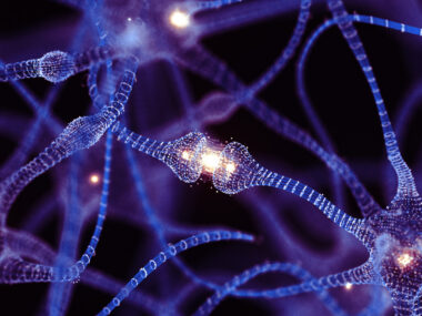
Researchers have created a detailed map of the mouse nervous system, including the location of the many diverse cell types in the brain, in the largest study yet of the makeup of the mammalian nervous system.
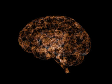
The new map could provide new clues about the origin of neurological diseases such as multiple sclerosis (MS). Researchers are now planning to use the same methods to build a detailed map of the human brain.
Findings were published in the study, “Molecular Architecture of the Mouse Nervous System,” was published in the journal Cell.
To map out in detail the diversity of cells within the nervous system, researchers used a new technology called single-cell RNA sequencing, which measures the specific expression of genes in single cells. Gene expression is the process by which information in a gene is synthesized to create a working product, such as a protein.
By measuring the gene activity of individual cells from well-defined regions within the nervous system, Karolinska Institutet researchers created a profile for each cell type and mapped them to specific regions, more systematically and more in depth than ever before.
“You can compare it to the difference between a medieval map and a satellite image: thousands of details that were previously invisible become visible with the use of these new techniques, and the entire map becomes more reliable,” Sten Linnarsson, PhD, a professor in the Department of Medical Biochemistry and Biophysics at Karolinska Institute, said in a press release.
The nervous system is composed of a wide variety of cells, one of the most important being glia cells, which include a broad range of cells (e.g., astrocytes, oligodendrocytes, and microglia) that have essential supportive and protective roles and, in fact, outnumber nerve cells. They can also produce myelin, the fat-rich substance that surrounds nerve fibers, and that is progressively destroyed in MS.
A number of cells from the immune system also exist in the nervous system, surveilling and protecting it against harmful invaders (bacteria, viruses, etc). Cells that make up vessels and membranes also coexist within the nervous system.
Using single-cell RNA sequencing, researchers found previously unknown cell types, determining their location with high precision.
Cells were mapped from several mouse tissues including the brain and spinal cord (central nervous system), ganglia surrounding the spinal cord (peripheral nervous system), and the nervous system that controls the gut (enteric nervous system).
The team identified 265 different types of cells, and found that neurons had the greatest diversity with more than 200 different types.
One of the most surprising discoveries was that astrocytes are more diverse than previously thought, and likely play distinct roles in different parts of the brain. Astrocytes are star-shaped cells that have multiple functions including providing nutrients to the nervous system and aiding in the repair and scarring process of the brain and spinal cord after traumatic injuries.
These findings can be used to understand the origin of different neurological diseases. Conditions such as MS are thought to originate from a specific type of cell, in a specific location and at a specific time depending on where and when relevant genes are expressed.
“With the help of our new atlas of the nervous system, researchers are now able to place disease-causing genes in specific cell types, which provides us with clues as to how the disease occurs,” Linnarsson said. “In the long run this might contribute to the development of new drugs or other therapies.”
Using the mouse model as a starting point, researchers now plan to uncover the architecture of the human brain using the same approach.

LAURIE HYTNER
I don't want new drugs. I want a cure.