
Infection with Common Herpes Virus Speeds MS-like Disease Onset and Progression in Primate Model, Study Reports
Written by |
Infection with the most common member of the herpes virus family, called HHV-6, may pass unnoticed and without symptoms, but the very act of being infected significantly accelerated the development and progression of a multiple sclerosis-like disease in nonhuman primates, a study reports.
Its findings support the role of viral infection in triggering “inflammatory-mediated neurologic conditions” like MS, it concluded.
The study, “Herpesvirus trigger accelerates neuroinflammation in a nonhuman primate model of multiple sclerosis,” was published in the journal Proceedings of the National Academy of Sciences of the United States of America (PNAS).
Several herpes viruses — including HHV-6, one of the most common and estimated to infect 80 percent of people during childhood — are linked with MS.
Specifically, a previous study reported that HHV-6 is found at increased frequency in MS lesions and outside the brain (for example, in circulating blood) in periods of MS clinical exacerbation. Another study suggested that dormant HHV-6 can hinder the repair mechanisms of the myelin sheath (the protective coat surrounding neurons), and the loss of myelin is the underlying cause of MS.
Since HHV-6 is almost always acquired in early childhood, it is difficult to assess its role in MS, a disease that usually manifests in early adulthood.
Join the MS forums: an online community especially for patients with MS.
Mice and other rodents are not susceptible to HHV-6 infection. So to study how HHV-6 infection affects MS onset, researchers at the National Institute of Neurological Disorders and Stroke (NINDS) used a monkey species, called marmosets, as animal models.
“Marmosets are excellent models of experimental autoimmune encephalomyelitis (EAE),” the researchers wrote, noting they better captures features of human MS than do rodents.
Researchers gave the marmosets intranasal administrations of HHV-6 — they injected separately two virus strains, HHV-6A or HHV-6B – or a solution in a control group for four months. The injections led to no clinical symptoms.
They then checked whether the animals mounted an immune response against the virus, and found those given the HHV-6 virus were positive for antibodies against it, while control animals were negative.
Whole brain analysis of one infected marmoset —the animal had died due to a wasting disease common in captive marmosets — showed signs of the presence of HHV-6.
Next, researchers set out to study how a pre-existing HHV-6 viral infection might affects EAE progression. They induced EAE in both infected and control animals, an monitored them frequently for signs of disease.
In marmosets given HHV-6, the onset of EAE symptoms was significantly quicker than in controls — the HHV-6 animals developed symptoms around day 80 after EAE induction, while controls showed no symptoms at day 200.
EAE marmosets infected with HHV-6 — either HHV-6A or HHV-6B — also died in a significantly shorter period following EAE induction compared to control animals.
Brain scans using magnetic resonance imaging (MRI), performed every two weeks after EAE induction, showed a trend for HHV-6-infected animals to develop lesions earlier than controls.
At the study’s end, determined as the time when all animals showed signs requiring them to be euthanized, researchers analyzed samples of their central nervous system (CNS, brain and spinal cord). Once again, they detected signs of HHV-6 in the virus-inoculated animals in several locations within the CNS. In the brain of a subgroup of animals infected with HHV-6 but without induced EAE, evidence of the virus was rare. Control animals were negative for HHV-6.
The HHV-6–positive brain lesions were also positive for immune cell infiltrates, namely with lymphocytes (a type of white blood cell).
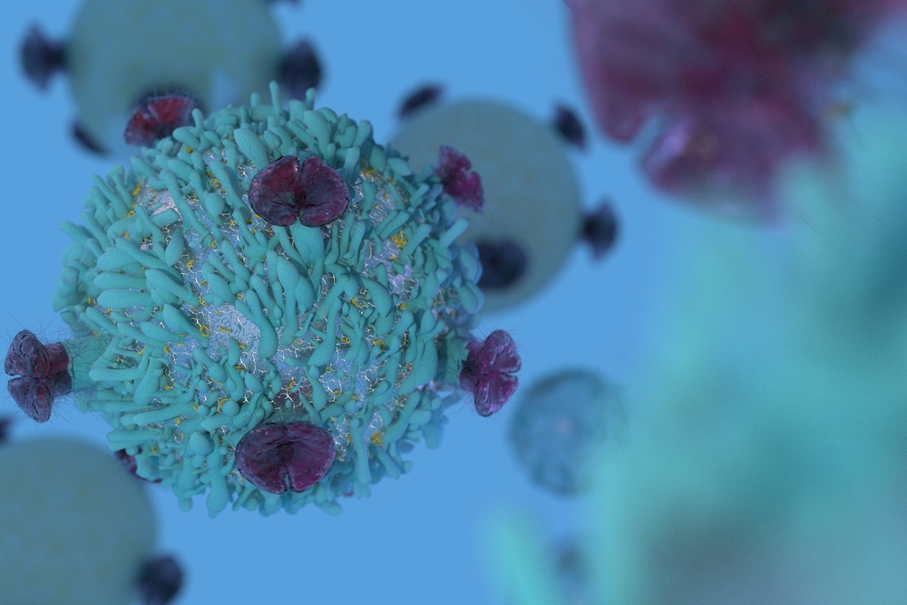
Finally, researchers found greater numbers of a subset of proinflammatory immune cells called CD8 T-cells — cells that secrete a proinflammatory signaling molecule called interferon gamma — in the EAE marmosets infected with HHV-6.
“Our data support the hypothesis that viruses may act as triggers to lower the threshold for autoimmunity, and warrant trials of antiviral interventions in early disease stages,” the researchers wrote.
Their findings, they concluded, support “the long- standing idea that virus(es) may act as triggers in MS or other inflammatory-mediated neurologic conditions.”


Anna Maria Sifo
Me, a nobody, have always "thought" this be the most reasonable theory, explaining MS in children. Might be a brain, marrow or eye herpes lurking . Acyclovir trials...
Sarah
I suffer re-occurring Herpes Simplex which developed years after my first MS relapse but the MS has progressed quicker since developing Herpesvirus. I am sure they will discover that it's with all herpesviruses, I always thought that there was a link.
Martin Matko
Thought you should find this interesting!
https://www.facebook.com/groups/191328180934162/permalink/1931750616891901/