ACTRIMS 2023: Portable MRI device able to detect brain lesions in MS
The imaging tool, the size of the R2-D2 droid, could go to patients
Written by |

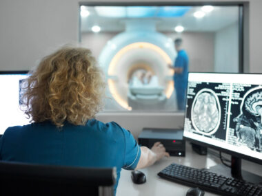
A portable MRI device may be used to detect brain lesions that are indicative of multiple sclerosis (MS), and serve as a low-cost imaging tool that can be brought to patients for testing, according to a new analysis.
While the system is not yet accurate enough to be used to diagnose the neurodegenerative disease itself, efforts are underway to improve it. The hope is that this portable MRI system ultimately could prove a useful diagnostic tool for MS in the future.
The device is about the size of R2-D2, the famous droid from “Star Wars,” according to researchers.
Serhat Okar, MD, a neurologist and post-doctoral research fellow at the National Institute of Neurological Disorders and Stroke (NINDS), part of the National Institutes of Health, presented the analysis’ findings at the Americas Committee for Treatment and Research in Multiple Sclerosis (ACTRIMS) Forum, being held in San Diego, California.
Okar’s talk was titled “Sensitivity of Portable Ultra Low Field Magnetic Resonance Imaging for Radiological Diagnosis of Multiple Sclerosis.“

Goal is a portable MRI that can diagnose MS
MS is caused by inflammation in the central nervous system, comprised of the brain and spinal cord, that results in lesions — areas of damaged and scarred tissue. Detecting lesions is crucial for diagnosing MS as, according to current diagnostic criteria, lesions must be present in multiple regions of the brain and/or spinal cord in order to confirm a formal diagnosis.
Magnetic resonance imaging, or MRI, is the gold standard for detecting MS lesions. MRI uses magnetic fields combined with radio waves to image the body’s internal structures.
Most MRI machines use large magnets to generate high-powered magnetic fields — generally, more powerful magnets result in clearer images, allowing more precise identification of lesions. However, use of these large magnets means that MRI machines usually are only available in a hospital; they cannot be used in a point-of-care approach where clinicians go to patients, rather than having patients come to a hospital.
Scientists have recently developed a new form of MRI machine that uses a low-powered magnet and is small enough to be portable — according to Okar, this novel device is about the size of the science fiction robot R2-D2, made famous in the “Star Wars” movies.
This ultra-low-field MRI (ULF-MRI) technology could theoretically be helpful for diagnosing and screening for MS in a point-of-care setting, assuming it can detect lesions with similar accuracy to standard MRI scans.
To test the accuracy of the system, a team led by researchers at NINDS conducted a small study that included 55 adults with MS. The average age of the participants was 51, and 43 of them were women. All of the patients underwent an MRI using a standard machine and a scan using ULF-MRI; both scans were conducted on the same day for each participant.
Two expert radiologists reviewed the scans obtained via ULF-MRI to identify lesions. The lesions identified on ULF-MRI were then compared with data from standard MRI scans to assess the accuracy of the detected lesions.
The results showed some variation between the two radiologists who reviewed the data. One of the experts tended to be more conservative in judging lesions, resulting in many missed lesions but also few false-positives. The other expert tended to be more liberal, which led to the correct identification of more lesions, but also a higher rate of false-positives.
Closer inspection of the ULF-MRI images and comparison with images from standard MRI scans showed that certain kinds of false-positives were especially common on images acquired with the portable machine. For example, ULF-MRI images often erroneously appeared to show lesions in the more densely folded regions of the brain.
In other regions — particularly in the area surrounding fluid-filled cavities in the brain called the periventricular region — ULF-MRI showed high accuracy for detection. This highlights the new system’s potential as a tool for MS diagnosis, Okar said.

Testing the portable MRI for accuracy, false-positives
After the initial round of judging lesions, the two experts were given side-by-side comparisons of ULF-MRI and standard MRI images from 10 of the patients, providing them an opportunity to learn from their mistakes and become more familiar with the common false-positives.
The experts then re-assessed ULF-MRI scans for 10 other patients. Both radiologists tended to get more conservative after the review opportunity, marking fewer lesions overall. This resulted in a modest decline in the number of correctly identified lesions, but almost completely eliminated false-positives.
Okar said that, overall, given the presence of false-positives and commonly missed regions, “radiological [diagnostic] criteria for MS may not be applicable to the portable ULF-MRI assessments at this point.”
Nonetheless, Okar said he’s optimistic that, with further improvements and refining, the ULF-MRI system could become a useful diagnostic tool, and also help monitor patients for relapses and measures of brain volume.
Efforts are underway to improve the system, including work to enhance the clarity of the images obtained. As a step toward implementing the system in routine practice, the researchers also are working to create a mobile MRI unit.
Note: The Multiple Sclerosis News Today team is providing in-depth coverage of the ACTRIMS Forum 2023 Feb. 23–25. Go here to see the latest stories from the conference. Follow along on Facebook, Twitter, and Instagram for live updates using the hashtag #actrims2023.