Researchers Report Encouraging Results in Mouse Model MS Stem Cell Therapy Research
Written by |
Scientists at the Scripps Research Institute (TSRI), the University of California (UC), Irvine and The University of Utah report that mice crippled by an autoimmune disease similar to multiple sclerosis (MS) regained the ability to walk and run after a team of researchers implanted human stem cells into their injured spinal cords, and remarkably — the mice recovered even after their bodies rejected the human stem cells.
 “When we implanted the human cells into mice that were paralyzed, they got up and started walking a couple of weeks later, and they completely recovered over the next several months,” says study co-leader Jeanne Loring, a professor of developmental neurobiology at TSRI, and team leader at the https://www.scripps.edu/loring/ the Loring Lab in the Sanford Consortium for Regenerative Medicine in La Jolla, California., whose mission is to advance stem cell research through collaborative, multi-disciplinary interactions. This “collaboratory” enables scientists from the La Jolla Institute for Allergy & Immunology, the Salk Institute for Biological Studies, the Sanford-Burnham Medical Research Institute, The Scripps Research Institute, and the University of California, San Diego to work side by side in a facility specifically designed to achieve breakthrough discoveries. Consistent with the objectives of the California Institute for Regenerative Medicine, Sanford Consortium researchers are applying the powers of stem cells to promote diagnoses, treatments, and cures for degenerative diseases and injuries.
 Dr. Thomas Lane, an immunologist and University of Utah Professor of Pathology who co-led the study with Dr. Loring, says he had never seen anything like it. “We’ve been studying mouse stem cells for a long time, but we never saw the clinical improvement that occurred with the human cells that Dr. Loring’s lab provided,” observes Dr. Lane, who began the study at UC Irvine, in a TSRI release.
Dr. Tom Lane’s laboratory has a long-standing interest in understanding events that initiate and maintain inflammation within the CNS in response to viral infection. divided into two main research objectives: 1) define the functional contributions of chemokines and chemokine receptors in defense and disease following viral infection of the central nervous system (CNS) and 2) evaluate the therapeutic potential of mouse/human neural precursor cells (NPCs) in clinical recovery and remyelination in a model of viral-induced demyelination.
Following instillation of a positive-strand RNA virus (mouse hepatitis virus – MHV) into the CNS of susceptible mice, the researchers have shown that blocking chemokine function via both antibody neutralization and genetic silencing in virally-infected mice resulted in increased mortality accompanied by reduced immune cell infiltration into the CNS, and have subsequently demonstrated that unique chemokine/chemokine receptor signaling pathways are critical for interrelated events required for optimal host defense following viral infection including linking innate and adaptive immune responses, regulating antiviral effector functions e.g. cytokine secretion/cytolytic activity by effector T cells, and promoting the directional migration of antigen-sensitized lymphocytes into the CNS.
They note that MHV infection of the CNS results in viral persistence in white matter tracts leading to chronic infiltration of activated lymphocytes and macrophages that contribute to demyelination. Importantly, generation of autoreactive T lymphocytes specific for myelin proteins e.g. myelin basic protein (MBP) and proteolipid protein (PLP) is not prevalent therefore epitope-spreading is not a relevant aspect in this model system. Additionally, MHV-induced demyelination results in moderate-to-severe clinical disease characterized by hind-limb paralysis.
Similar to the human demyelinating disease multiple sclerosis (MS), remyelination failure is also observed in MHV-infected mice. Consequently, an important and clinically-relevant question related to demyelinating diseases is to design therapies that promote remyelination of demyelinated axons. In the lab Dr. Lane’s team have previously shown that surgical engraftment of syngeneic neural progenitor cells (NPCs) into mice persistently-infected with MHV results in survival and migration of engrafted NPCs accompanied by extensive remyelination.
“We are the only group, to my knowledge,” says Dr. Lane, “examining the therapeutic potential of cell replacement strategies using a viral model of demyelination. This is important in that the etiology of MS remains enigmatic and viruses have long been considered important as a potential triggering agent in inducing demyelinating diseases such as MS. Moreover, numerous viruses are capable of persisting within the CNS therefore understanding if NPCs are capable of promoting repair within this environment is critical. We have determined that transplanted cells migrate to areas of demyelination by responding to the specific chemokines expressed within areas of demyelination. We have now moved forward with our studies on NPC-mediated clinical/histological recovery to address whether allogeneic NPCs are antigenic and subject to immune-mediated rejection. We are also investigating the therapeutic potential of human NPCs (hNPCs) in mediating functional recovery following transplantation into MHV-infected mice. We have determined that intraspinal transplantation of hNPCs into MHV-infected mice results in rapid rejection. Therefore, we are now interrogating mechanisms by which to prolong hNPC survival. More importantly, we have shown that transplantation of hNPCs results in sustained clinical recovery lasting out to 6 months post-transplant (p.t.). Our data indicates that hNPCs are immunosuppressive as evidenced by dramatic reduction in neuroinflammation accompanied by remyelination within the spinal cords of transplanted animals. These findings are provocative and indicate that hNPCs are indeed therapeutic although rapidly rejected.”
[adrotate group=”4″]
Dr. Lane notes that the remyelination failure observed in multiple sclerosis is also observed in MHV-infected mice. Consequently, an important and clinically-relevant question related to demyelinating diseases is to design therapies that promote remyelination of demyelinated axons. The team have determined that surgical engraftment of mouse neural precursosr cells (NPCs) into mice persistently-infected with MHV results in survival and migration of engrafted NPCs accompanied by extensive remyelination.
Stem Cell Therapy for MS
In MS, immune cells known as T cells invade the upper spinal cord and brain, causing inflammation and ultimately the loss of an insulating coating on nerve fibers called myelin. Affected nerve fibers lose their ability to transmit electrical signals efficiently, and this can eventually lead to symptoms such as limb weakness, numbness and tingling, fatigue, vision problems, slurred speech, memory difficulties and depression.
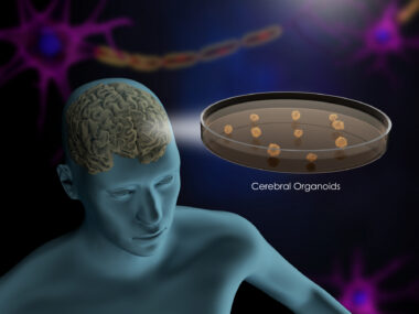
Current MS therapies, such as interferon beta, aim to suppress the immune attack that strips the myelin from nerve fibers. But they are only partially effective and often have significant adverse side effects. Dr. Loring’s group at TSRI has been searching for another way to treat MS using human pluripotent stem cells, which are cells that have the potential to transform into any of the cell types in the body.
The research team has been focused on turning human stem cells into neural precursor cells, which are an intermediate cell type that can eventually develop into neurons and other kinds of cells in the nervous system. In collaboration with Dr. Lane’s group, Dr. Loring’s team has been testing the effects of implanting human neural precursor cells into the spinal cords of mice that have been infected with a virus that induces symptoms of MS.
Dr. Loring’s group has been focused on turning human stem cells into neural precursor cells, which are an intermediate cell type that can eventually develop into neurons and other kinds of cells in the nervous system. In collaboration with Lane’s group, the Loring team has been testing the effects of implanting human neural precursor cells into the spinal cords of mice that have been infected with a virus that induces symptoms of MS.
The Loring Lab employs a broad spectrum of basic and translational research approaches to investigate the dynamic nature of stem cells conducted by a team of highly motivated researchers, and the lab has gained a reputation in the stem cell community for its experimental rigor and solid contributions to the scientific literature, a status it maintains by staying on the brink of experimental innovation by incorporating some of the most cutting edge technologies in the life sciences. The lab’s primary goals are to bridge the gap between understanding stem cells at a molecular level and applying that knowledge to the field of regenerative medicine.
In an Open Access article published online ahead of print by the journal Stem Cell Reports, entitled “Human Neural Precursor Cells Promote Neurologic Recovery in a Viral Model of Multiple Sclerosis” (Published Online: May 1, 2014, DOI: https://dx.doi.org/10.1016/j.stemcr.2014.04.005), Drs. Walsh, Lane, and Loring along with TSRI colleagues Ron Coleman, and Ha Tran, and co-authors Lu Chen, Ronika Leang, Alexandra Kopf, Ilse Sears-Kraxberger and Oswald Steward of UC Irvine, and Wendy B. Macklin of the University of Colorado School of Medicine, describe how by using a viral model of multiple sclerosis, they show that intraspinal transplantation of human embryonic stem cell-derived neural precursor cells (hNPCs) results in sustained clinical recovery, even though hNPCs were not detectable beyond day eight posttransplantation.
They report that Improved motor skills were associated with a reduction in neuroinflammation, decreased demyelination, and enhanced remyelination, and that evidence indicates that the reduced neuroinflammation is correlated with an increased number of CD4+CD25+FOXP3+ regulatory T cells (Tregs) within the spinal cords. Coculture of hNPCs with activated T cells resulted in reduced T cell proliferation and increased Treg numbers. The hNPCs acted, in part, through secretion of TGF-1 and TGF-2. They conclude that these findings indicate that the transient presence of hNPCs transplanted in an animal model of MS has powerful immunomodulatory effects and mediates recovery, and that further investigation of the restorative effects of hNPC transplantation may aid in the development of clinically relevant MS treatments.
 The mice’s dramatic recovery after human stem cells were implanted, could lead to new ways to treat multiple sclerosis in humans. “This is a great step forward in the development of new therapies for stopping disease progression and promoting repair for MS patients,” says study co-author Craig Walsh, a UC Irvine immunologist and Associate Professor of Molecular Biology and Biochemistry, and Associate Director of the Institute for Immunology in the School of Biological Sciences.
Current MS therapies, such as interferon beta, aim to suppress the immune attack that strips the myelin from nerve fibers, but they are only partially effective and often have significant adverse side effects. Dr. Loring’s group at TSRI has been searching for an alternative way to treat MS using human pluripotent stem cells, which are cells that have the potential to transform into any of the cell types in the body. Dr. Loring’s group has been focused on turning human stem cells into neural precursor cells, which are an intermediate cell type that can eventually develop into neurons and other kinds of cells in the nervous system. In collaboration with Dr. Lane’s group, Dr. Loring’s team has been testing the effects of implanting human neural precursor cells into the spinal cords of mice that have been infected with a virus that induces symptoms of MS.
The transformation that took place in the largely immobilized mice after human neural precursor cells were injected into the animals’ damaged spinal cords was dramatic, says Dr. Loring in the TSRI release. “Tom called me up and said, ‘You’re not going to believe this’. He sent me a video, and it showed the mice running around the cages. I said, ‘Are you sure these are the same mice?’”
 Even more remarkable, as noted above, the animals continued walking even after the human cells were rejected, which occurred about a week after implantation. This suggests that the human stem cells were secreting a protein or proteins that had an enduring effect on preventing or impeding progression of MS in the mice comments Ron Coleman, a TSRI graduate student in Dr. Loring’s lab who was co-first author of the Cell paper with Lu Chen of UC Irvine. “Once the human stem cells kick that first domino, the cells can be removed and the process will go on because they’ve initiated a cascade of events,” says Mr. Coleman.
The scientists showed in the Cell study that the implanted human stem cells triggered the creation of white blood cells known as regulatory T cells, which are responsible for shutting down the autoimmune response at the end of an inflammation. In addition, the implanted cells released proteins that signaled cells to re-myelinate the nerve cells that had been stripped of their protective sheaths.
The particular line of human neural precursor cells used to heal the mice was the result of a lucky break. Coleman was using a common technique for coaxing human stem cells into neural precursor cells, but decided partway through the process to deviate from the standard protocol. In particular, he transferred the developing cells to another Petri dish.
“I wanted the cells to all have similar properties, and they looked really different when I didn’t transfer them,” says Mr. Coleman, who was motivated to study MS after his mother died from the disease. This step, called “passaging,” proved key. “It turns out that passaging alters the types of proteins that the cells express,” he says.
A Happy Accident
Dr. Loring calls the creation of the successful neural precursor cell line a “happy accident.” “If we had used common techniques to create the cells, they wouldn’t have worked,” she said. “We’ve shown that now. There are a dozen different ways to make neural precursor cells, and only this one has worked so far. We now know that it is incredibly important to make the cells the same way every time.”
The research team is now working to discover the particular proteins that its unique line of human precursor cells release. One promising candidate is a class of proteins known as transforming growth factor beta, or TGF-B, which other studies have shown is involved the creation of regulatory T cells. Experiments by the scientists showed that the human neural precursor cells released TGF-B proteins while they were inside the spinal cords of the impaired mice. However, it’s also likely that other, as yet unidentified, protein factors may also be involved in the mice’s healing.
If the team can pinpoint which proteins released by the neural precursor cells are responsible for the animals’ recovery, it may be possible to devise MS treatments that don’t involve the use of human stem cells. “Once we identify the factors that are responsible for healing, we could make a drug out of them,” says Dr. Lane. Another possibility, Dr. Loring notes, might be to infuse the spinal cords of humans affected by MS with the protein factors that promote healing.
A better understanding of what makes these human neural precursor cells effective in mice will be key to developing either of these therapies for humans. “We’re on the trail now of what these cells do and how they work,” Dr. Loring affirms.
The Scripps Research Institute (TSRI) is one of the world’s largest independent, not-for-profit organizations focusing on research in the biomedical sciences, and is internationally recognized for its contributions to science and health, including its role in laying the foundation for new treatments for cancer, rheumatoid arthritis, hemophilia, and other diseases. An institution that evolved from the Scripps Metabolic Clinic founded by philanthropist Ellen Browning Scripps in 1924, the institute now employs about 3,000 people on its campuses in La Jolla, CA, and Jupiter, FL, where its renowned scientists – including three Nobel laureates – work toward their next discoveries. The institute’s graduate program, which awards PhD degrees in biology and chemistry, ranks among the top ten of its kind in the nation.

The Division of Microbiology and Immunology (M&I) in the University of Utah School of Medicine Department of Pathology was established in the early 1970s by past chairman Ernst Eichwald, M.D. Dr. Eichwald, an internationally recognized clinician/scientist who recruited a group of faculty to Utah with shared interests in better understanding the immunological mechanisms underlying graft infection and transplantation tolerance.
Sources:
The Scripps Research Institute
The University of California (UC), Irvine
The University of Utah
Stem Cell Reports
Image Credits:
The Scripps Research Institute
The University of California (UC), Irvine
The University of Utah
Stem Cell Reports