Better MS Tracking Tool Developed by Robarts Institute Scientists At University of Western Ontario
Written by |

A team of magnetic imaging scientists led by Dr. Ravi Menon, PhD, at the University of Western Ontario’s Robarts Research Institute have developed a better way to track the progression of Multiple Sclerosis (MS) from its very early stages. The Robarts researchers used a technique called Quantitative Susceptibility (QS) Magnetic Resonance Imaging (MRI), to measure a sort of damage in specific areas of the brain which the study showed to be common to all patients. The findings are published in advance online, in the journal Radiology.
Entitled “Multiple Sclerosis: Improved Identification of Disease-relevant Changes in Gray and White Matter Using Susceptibility-based MR Imaging” (DOI: https://dx.doi.org/10.1148/radiol.14132475) the paper is coauthored by Dr. Menon with David A. Rudko , PhD; Igor Solovey , MSc; Joseph S. Gati , MSc; and Marcelo Kremenchutzky , MD variously associated with the Department of Physics (D.A.R., R.S.M.), Center for Functional and Metabolic Mapping, Robarts Research Institute (D.A.R., I.S., J.S.G., R.S.M.), and the Department of Neurology, University Hospital (M.K.), University of Western Ontario London, Ontario, Canada.
The coauthors describe their research as an evaluation of the potential of quantitative susceptibility (QS) and R2* mapping as surrogate biomarkers of clinically relevant, age-adjusted demyelination and iron deposition in multiple sclerosis. All study participants gave written informed consent, and the study was approved by the institutional review board. Quantitative maps of the magnetic resonance imaging susceptibility parameters (R2* and QS) were computed for 25 patients with either clinically isolated syndrome (CIS) or relapsing-remitting MS, as well as for 15 age and sex-matched control subjects imaged at 7 T. The candidate MR imaging biomarkers were correlated with Extended Disability Status Scale (EDSS), time since CIS diagnosis, time since MS diagnosis, and age.
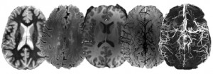
The process employed used a standard Siemens 3T MRI so it could be reproduced in any hospital, using the technique, called QS. The researchers mapped this MRI parameter in 25 patients with relapsing-remitting MS or clinically isolated syndrome (CIS half of those diagnosed with CIS will go on to be diagnosed with MS) measuring both demyelination and iron deposition. Fifteen age and sex-matched control subjects were also scanned. While brain and spine lesions visualized with normal MRI tend to appear and disappear over time, QS shows common areas of damage in all patients that correlated very well with the Extended Disability Status Score (EDSS) which is a standard tool used to measure MS progression, as well as with age and time since diagnosis.
The researchers report that QS maps aided identification of significant, voxel-level increases in iron deposition in subcortical gray matter (GM) of patients with MS compared with control subjects, and that Voxelwise QS also supported a significant contribution of age to demyelination in patients with MS, suggesting that age-adjusted clinical scores may provide more robust measures of MS disease severity compared with non age-adjusted scores. They conclude that using QS and R2* mapping, evidence of both significant increases in iron deposition in subcortical GM and myelin degeneration along the WM skeleton of patients with MS was identified. Both effects correlated strongly with EDSS.
“In MS research, there is something we call a clinical-radiological paradox,” says Dr. Menon in a Robarts Institute release. “When you do conventional MRIs on these patients you see lesions in the brain very clearly, but the number or volume of their lesions do not correlate with the patients disabilities. This paradox has been recognized since the MRI was introduced to clinical practice in the early 80s, and yet this is the only imaging tool we have for assessing MS. Our research provides a quantitative tool using a relatively conventional imaging sequence but with novel analysis. This tool shows that there is considerable damage occurring in common areas of all patients in both the white matter and in the deep brain structures the gray matter. Those quantitative measures what we call quantitative susceptibility, correlate with disease symptoms.”
[adrotate group=”4″]
“Significantly, in white matter, even where we see no lesions whatsoever, we’re able to measure damage in the same area of all patients using QS mapping. So even at the very earliest stages of the disease when the disability score is very low, or when the person hasn’t yet been diagnosed with MS, theres already significant damage,” added Dr. Menon, who holds the Canada Research Chair in Functional and Metabolic Mapping, and is a Professor of Medical Biophysics, Diagnostic Radiology & Nuclear Medicine, Neuroscience, Biomedical Engineering, and Psychiatry At UWO’s Robarts Research Institute. “This could have important diagnostic and prognostic implications, as there are drugs available to slow or stop the progression of MS, if started early enough.”
The Robarts Research Institute’s Centre for Functional and Metabolic Mapping (CFMM) at UWO houses Canada’s only collection of high-field (3T human) and ultra-high field (7T human and 9.4T animal) MR systems.
The Centre is dedicated to establishing the anatomical, metabolic and functional characteristics of normal brain development and healthy aging across the lifespan; as well as establishing the brain basis of developmental, neuropsychiatric and neurodegenerative deficits. The Laboratory for Functional Magnetic Resonance Research (LfMRR) is located in a purpose built addition to the Robarts Research Institute on the UWO Campus.
Dr. Menon’s major research focus is in the field of functional magnetic resonance imaging (fMRI), a method that allows neuroscientists to examine brain function at high spatial and temporal resolution. Another major area is in the design of sophisticated Radio Frequency hardware to better utilize these ultra-high field magnets, as well as the development and application of magnetic resonance imaging (MRI) and spectroscopy (MRS) techniques. MRI and MRS offer powerful methods for the non-invasive evaluation of the brain in health and disease using the 3T Siemens Tim Trio and the only 7T Varian/Siemens magnetom 7!Tesla MRI scanner in Canada (both human) and the 9.4T Varian (preclinical) MRI systems in the Centre for Functional and Metabolic Mapping.
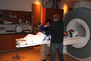
 The MAGNETOM Tim Trio whole-body 3T MRI scanner is currently the workhorse of the Centre for Functional and Metabolic Mapping, where at any given time, approximately 30 fMRI, spectroscopy, and diffusion studies underway on the 3T system will be underway.
The 3T Tim Trio is a short-bore magnet, with a 60cm bore diameter. The system contains 102 seamlessly integrated coil elements and 32 RF channels, with the ability to scan the full body with a field of view of 50 cm and up to 181 cm in length. The maximum gradient strength is 45 mT/m with a slew rate up to 200 T/m/s, and both active and passive noise reduction technology greatly reduces noise level compared to traditional systems. The integrated Tim (Total imaging matrix) technology provides unprecedented flexibility, accuracy, and speed, and is capable of reducing scan times by up to 50%.
The LfMRR also houses Canada’s highest field (4 Tesla) Magnetic Resonance Imaging / Magnetic Resonance Spectroscopy (MRI/MRS) scanner. The 4T facility is used primarily for in-vivo studies of human brain structure and function. These studies include assessment of brain metabolism and physiology, cognitive function and vascular dynamics, not only in normal and research patient populations, but also in in-vitro and animal models using a variety of advanced nuclear magnetic resonance imaging and spectroscopy techniques. A magnetic field strength of 4 Tesla (4T) is currently the highest field approved for human use. The 4T facility represents a unique national resource for state-of-the-art evaluation of brain structure and functional activity using a variety of MRI and MRS.
“My research area lies in the development of technology for ultra high field functional magnetic resonance imaging (fMRI) as well as understanding what it measures and tells us about human brain function, comments Dr Menon. Driven by neuroscience questions posed by our colleagues, we develop new hardware (such as new radio frequency coils) and software solutions (novel pulse sequences) for fMRI. We utilize MRI scanners at 3 Tesla (T), 7 T and 9.4 T to understand the normal anatomy and function of both humans and animals as well as to understand the changes in anatomy and function that accompany disease and degeneration in the brain. These studies are done with colleagues in the hospitals and University, most notably in the Centre for Brain and Mind.”
Dr. Menon describes the work his department at Robarts is doing in a collection of videos that can be accessed here:
https://cfmm.robarts.ca/media/
The research study “Multiple Sclerosis: Improved Identification of Disease-relevant Changes in Gray and White Matter Using Susceptibility-based MR Imaging” was funded by the Canadian Institutes of Health Research.
Sources:
University of Western Ontario
Robarts Research Institute
Centre for Functional and Metabolic Mapping
Radiology
Image credits:
Robarts Research Institute
Centre for Functional and Metabolic Mapping


