
Leptomeningeal Inflammation May Offer New Treatment Targets In Progressive Forms of MS
Written by |
Researchers at Johns Hopkins University in Baltimore presented key findings today, Feb. 19, concerning the presence of contrast-enhancing lesions in later stages in the relapsing-remitting experimental autoimmune encephalitis (EAE) model. The presentation was made at the Americas Committee for Treatment and Research in Multiple Sclerosis (ACTRIMS) Forum 2016, which is ongoing through Feb. 20 in New Orleans, LA.
The presentation, “Leptomeningeal inflammation and Intrathecal Rituximab in Progressive MS,” was given by Dr. Peter Calabresi.
Progressive multiple sclerosis (MS), characterized by steady worsening of neurological functioning (without any distinct relapses, also known as attacks or exacerbations) still lacks effective treatments. An increasingly recognized feature, particularly with high prevalence in patients with primary or secondary progressive MS, is the presence of leptomeningeal inflammation (LMI).
The organs of the central nervous system, brain, and spinal cord, are covered by three connective tissue layers collectively called the meninges, and the leptomeninges are the two delicate layers composing the meninges. LMI is a condition associated with gray matter demyelination and increased sub-pial lesion load. Therefore, leptomeningeal inflammation may have a role in disease progression.
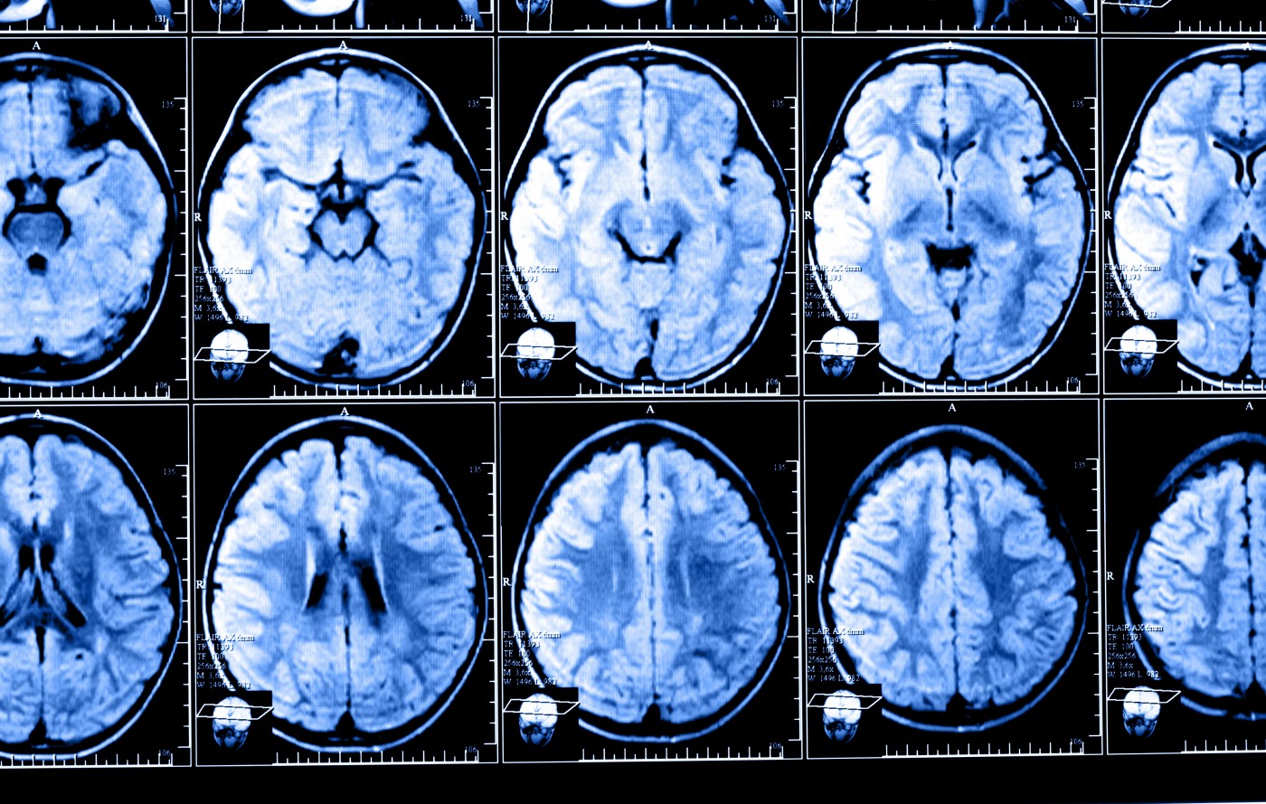
Capturing leptomeningeal lesions in vivo has been challenging, but a technique called time-delayed post-contrast fluid-attenuated inversion recovery (FLAIR), a pulse sequence used in magnetic resonance imaging (MRI), successfully identified these lesions, which were shown to correspond to inflammation foci composed by lymphocyte aggregates.
In the presentation, Calabresi showed the presence of contrast-enhancing lesions in a relapsing-remitting EAE model at later disease stages. Further studies confirmed that these lesions persisted over time and were composed by B- and T-lymphocytes. Thus, areas of inflammation with lymphocyte aggregates are also present in multiple sclerosis, suggesting a similar process to LMI observed in MS.
Treatments targeting LMI may have a potential therapeutic effect in MS. Currently, anti-CD20 is an approved treatment by the U.S. Food and Drug Administration (FDA), and it is administered intrathecally (injection into the spinal canal) to people with multiple sclerosis.
Based on this knowledge, the researchers initiated a clinical trial for Rituximab, a chimeric monoclonal antibody against the protein CD20, in patients with progressive MS who also have leptomeningeal enhancement, as confirmed by MRI.
The team plans to enroll 12 patients in total, and the goals of the trial are: to determine the safety of intrathecal administration of rituximab in patients with progressive MS; to investigate the effect of intrathecal rituximab on the persistence of leptomeningeal enhancement in patients with progressive MS; and to assess the effect of intrathecal rituximab on depleting cerebrospinal fluid (CSF) B-cells and lowering the levels of biomarkers of inflammation and neuronal damage.

Susan hoffman
Thank you for your work on the progressive form of MS.
Is there anything out there for reversing the damage done?
James Nielsen
I'll second that question !!!
We really not only need to stop disease progression, we also need repairative therapies...
Rebecca Bergeron
I'm in year 13 of a slow steady increase in symptoms. I've been dx w/ ms, then fibromyalgia, follows by lupus. Before I was even 2 yrs in I had pretty much every MS symptom on the list. These symptoms have never gone away and as stated, have been slowly and steady increasing. I tried copaxone for 5 yrs, then rebif for 8 months, before I stopped taking disease modifying drugs for awhile (per the neuro at Mayo, who verified that the previous treatments were ineffective.
Years later, I was talked into trying tysabri. The good news? It helped. It seems to have slowed down my progression. More than that, my cognitive ability has greatly improved (one of my worst symptoms), along w/mobility, and more. All the symptoms are still there, but it seems like since my body doesn't have to work so hard on the disease I'm able to work on rerouting my brain better.
On top of that, after onset in '03, I retired in '05, and made taking care of myself my new job. I have to say I've been pretty successful. I bought a three story house so I'd have to keep taking the stairs. It wasn't easy at first, but now I barely notice them. I'm not saying theyre not still challenging, only that I accept them as a challenge and not as a problem. Since starting tysabri, I've also taken to walking an hour/day every day. This is also not that easy, but having done it for 2 yrs straight (same route), I know the tricks to unlock my legs when they turn to concrete, or even how to shift myself to allow a spasm to pass.
I guess my point, that I got off of, is that I believe we can repair damage done, or at least possibly reroute our brains to create new pathways fo old abilities. In my experience, it's all about adapting. Finding new ways to keep function, challenging your balance, using mirrors for postural awareness. Keeping our brains thinking, singing, remembering, using biofeedback to learn how to manually control things ( like your bladder, swallowing, etc.).
Think of yourself like a stroke victim. They lose ability and then do a ton of pt and potentially recover. Why can't we?
Sorry for the long post. While I can think clearer now, I not good at keeping it short. :)
Best wishes to all, good luck, God bless, and keep moving!
Becky