
Immune Cells in Brain Seen to Promote Cognitive Impairment In MS Mouse Model
Written by |
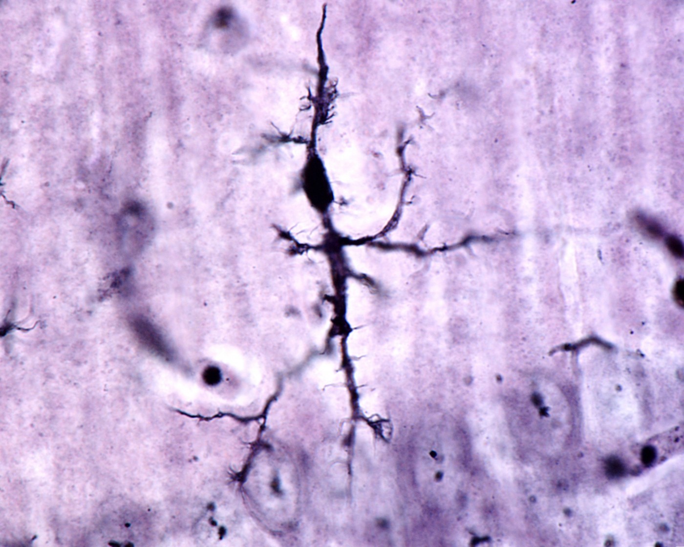
Microglial cells in the hippocampus, a brain region crucial for memory processing, may contribute to brain damage associated with cognitive impairment in multiple sclerosis (MS). The finding, published in the journal Scientific Reports, implies that targeting microglia could be a promising strategy to improve cognition in MS.
While cognitive impairment in MS is common, research has mainly focused on aspects of the disease related to demyelination. As a consequence, not much is known about the synaptic and molecular changes that contribute to a reduced cognitive performance.
Researchers at the University of Perugia, Italy, studied a mouse model of MS — experimental autoimmune encephalomyelitis (EAE) — that mimics the relapsing-remitting nature of one of the most common MS clinical variants. Specifically, they studied the hippocampus of mice in the remission phase of the disease, and found that microglial (immune cells of the brain and spinal cord) were also activated during remission, after motor symptoms had waned.
Long-term potentiation (LTP) is a measure of persistent strengthening of connections between neurons, called synapses. The study, “Persistent activation of microglia and NADPH drive hippocampal dysfunction in experimental multiple sclerosis,“ reported that this mechanism was reduced in the mice. Moreover, the EAE mice displayed problems in learning and recognition.
Activated microglia release a number of factors that are potentially toxic to neurons, such as pro-inflammatory cytokines and the enzyme NADPH oxidase, giving rise to reactive oxygen species. The researchers noted that NADPH oxidase was elevated in the mice, and was, in fact, mediating the effects of activated microglia on LTP. When the researchers blocked NADPH, the levels of LTP returned to normal.
IL-1b, a proinflammatory cytokine, was also increased in the mice. IL-1b is known to alter LTP in its own right, a finding also observed in the study. The LTP block was reversed when scientists blocked NADPH, showing that the effect of the cytokine is mediated by NADPH oxidase.
Finally, the team treated the mice with minocycline, a drug that prevents the activation of microglia. Minocycline prevented the release of NADPH oxidase and IL-1b, and restored both LTP and the performance in the cognitive test. However, the standard treatment used during MS relapses, methylprednisolone, had no effect on synaptic plasticity in the mice.
The report underscores the importance of further study into interactions between the immune and nervous systems during MS, and of exploring alternative treatment options targeting this interaction.
Shasha
Minocycline hurt me...hurt gut lining/lowered my immune system and hurt mitochondria. It caused strange cognitive errors since it can go into the brain/spinal cord. It is not help...hurt me. Raising oxygen in my brain helps my MS.