
How Astrocytes Promote Inflammation in Patients’ Brain Detailed in Early Study
Written by |
Using brain tissue from people with multiple sclerosis (MS) and mouse models of MS, scientists identified a key pathway that drives astrocytes to promote inflammation in the brain and spinal cord.
The study, “MAFG-driven astrocytes promote CNS inflammation” published in the journal Nature, uncovered potential therapeutic targets that may be tackled in future treatments to halt or slow MS progression.
“Astrocytes contribute to the pathogenesis [disease mechanisms] of multiple sclerosis, but little is known about the heterogeneity of astrocytes and its regulation,” the researchers wrote.
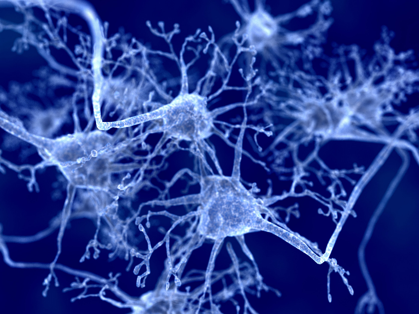
Star-shaped cells abundant in the central nervous system (CNS, the brain and spinal cord), astrocytes are involved in many activities that support CNS function and health. They provide nutrients to nerve cells (neurons), help repair injuries to nervous tissue, and regulate the transmission of nerve signals. They are believed to be the most abundant cells in the brain.
Despite their important contributions to the CNS, a population of neurotoxic astrocytes has been identified in several neurological diseases.
In MS, astrocytes are now recognized to be early and highly active players in the formation of demyelinating lesions. They seem to be key for recruiting immune cells to the brain and spinal cord, and for controlling immune cells that are active during progressive phases of MS.
They also react at a late stage by forming scar tissue once inflammation has subsided, a process that can prevent the repair of damaged myelin, the nerve-insulating sheath mistakenly attacked by the immune system in MS.
A team led by researchers at Harvard Medical School identified the molecules that drive astrocytes to promote inflammation, using MS mouse models and brain tissues collected from deceased MS patients and individuals without this disease.
The scientists used a novel technology known as single-cell RNA sequencing to track the activity, or expression, of genes in a single cell at a single point in time. As cells change over time, this tool can provide valuable insights into how specific cell populations contribute to disease processes.
Using this technology on both patient samples and mice with MS-like disease, the researchers identified astrocytes that had decreased production of NRF2 — a protein that protects against inflammation by switching on several genes associated with antioxidant pathways— while increasing the expression of MAFG — which cooperates with other proteins to repress antioxidant and anti-inflammatory effects by promoting DNA methylation, a type of DNA mark.
Results also provide evidence that inflammatory immune cells infiltrating the central nervous system during MS secrete a messenger called granulocyte-macrophage colony-stimulating factor (GM-CSF). This messenger amplifies the pro-inflammatory signals of astrocytes, boosting their contribution to CNS disease.
“These findings define previously undescribed mechanisms of disease pathogenesis, and identify … candidate targets to suppress the pathogenic [disease-causing] activity of astrocytes in MS,” the researchers wrote.
This research project was funded by the National MS Society and the International Progressive MS Alliance, among other institutions.
Of note, Francisco J. Quintana, PhD, the study’s senior author, was awarded the 2019 Barancik Prize for Innovation in MS Research for his work in understanding what causes MS, and in identifying potential therapeutic targets and biomarkers for the disease.


Leave a comment
Fill in the required fields to post. Your email address will not be published.