
#ACTRIMS2020 – CorTechs Says AI Models May Improve Early MS Diagnosis
Written by |
CorTechs Labs presented new data indicating that machine learning models based on magnetic resonance imaging (MRI) of the brain may aid in the early diagnosis of multiple sclerosis (MS).
Company representatives, joined by other experts, also released updated recommendations for a standardized MRI protocol for MS.
The findings and recommendations were presented at the Americas Committee for Treatment and Research in Multiple Sclerosis (ACTRIMS) Forum 2020, held Feb. 27-29 in Florida.
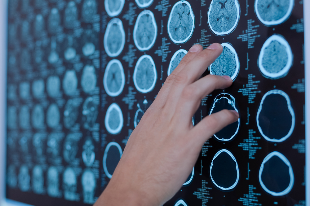
Early MS diagnosis is critical. Patients diagnosed early can start treatments sooner, which may prevent severe brain volume loss (brain atrophy), and physical and cognitive decline. Brain MRI has been an invaluable tool in diagnosis, key to accurate identification of new lesion activity.
Recent advances in image processing and machine learning have provided ways to deepen knowledge about MS, and gather new information that could improve its diagnosis.
In a poster titled “Performance Evaluation For Multiple Sclerosis Identification Models Based On MR Imaging And Machine Learning,” CorTechs demonstrated machine learning models for MS identification that were created using a recent version of its lead brain image software, NeuroQuant 3.0, which quantifies brain volume changes seen on MRI.
NeuroQuant-processed MRI scans from 463 MS patients and 2,315 healthy controls between the ages of 18 and 71 were used to train artificial intelligence (AI) algorithms to create the models.
Several types of volumetric information were used as input, including the volume of cerebral white matter hypointensities, a sign of brain lesions. Two-thirds of the data were used for training the algorithm, and one-third was used for testing the models.
Three different models were created, according to age.
Model one, incorporating all age groups, had a precision of 93% and a sensitivity of 100% for identifying healthy subjects, and a precision of 98% and sensitivity of 63% for identifying MS subjects. The sensitivity of a test indicates its ability to correctly identify those with the disease.
The main features influencing the model were normalized volumes of cerebral white matter hypointensities, the white matter of the thalamus and cerebellum (two brain regions), age, and the volume of isthmus cingulate (another brain region).
The second model — designed for subjects younger than 40 — made predictions with a precision of 92% for both healthy and MS subjects, and a sensitivity of 98% for healthy and 76% for MS subjects. Its three most important parameters were the same as in model one, plus two additional features with significant influence: normalized volumes of cerebral white matter and the posterior superior temporal sulcus (another brain region).
In the third model — for ages 40 and older — identification of healthy people and people with MS was possible with a precision of 94% and 96%, and sensitivity of 100% and 57%, respectively. Key parameters for that model included normalized volumes of the thalamus, age, cerebellar white matter, cerebral white matter hypointensities, and isthmus cingulate volume.
The models “might be used as MS classifiers to assist clinicians in decision-making and earlier MS diagnosis,” the researchers wrote, adding that the models showed “high classification performance” for distinguishing healthy people and people with MS.
Creating models adjusted for age “selected different key features, thus enabled different precision and sensitivity performance for MS classification,” they said.
In a second poster, “Updated Recommendations for a Standardized MRI Protocol for Multiple Sclerosis,” a panel of experts, including some from CorTechs, issued recommendations to update the Consortium of Multiple Sclerosis Centers (CMSC) MRI protocol, the standard MRI protocol used in North America for MS diagnosis and follow-up.
The goal is to promote the universal adoption of a standardized MRI protocol for MS.
The experts —neurologists, radiologists, and imaging scientists with expertise in MS — emphasized the “critical importance” of using the subcallosal plane and thin (less than or equal to 3 mm) image slices.
The suggested updated protocol includes a core analysis, mandatory for all MRI studies; a gadolinium analysis when indicated (gadolinium is a contrast agent used in MRI scans); and an optional analysis. It also includes a new category called optimum plus, consisting of 3D high-resolution images.
Other recommendations include surveillance for progressive multifocal encephalopathy, and judicious use of gadolinium.
The group also worked to approximate the CMSC protocol with the Magnetic Resonance Imaging in MS group (MAGNIMS) guidelines used in Europe, to reach international consensus.
“We are dedicated to improving patient outcomes and up-leveling the standard of patient care through best-in-class clinical applications and patient-centric analysis and reporting,” Chris Airriess, PhD, CorTechs Labs’ CEO said in a press release.
“Having two very close friends with the disease, I have seen firsthand the impact MS can have on the lives of patients and their families,” Airriess said. “We work hard every day to provide software solutions to enable early, accurate diagnosis of MS to ultimately help patients get the treatment they need as soon as possible —and to monitor treatment response to ensure the best possible outcomes. The research being presented at ACTRIMS provides hope for furthering these advancements in the diagnosis and classification of MS.”


Leave a comment
Fill in the required fields to post. Your email address will not be published.