
Immune T-cells Trigger Early MS Inflammation, Twin Study Reveals
Written by |
A comparison of immune cells isolated from identical twins — in which only one of each pair was diagnosed with multiple sclerosis (MS) — identified a population of immune-regulating T-cells present in those with asymptomatic brain inflammation, a study has found.
These findings suggest that T-cells may play a pivotal role in triggering MS.
The study, “Immune signatures of prodromal multiple sclerosis in monozygotic twins,” was published in the journal Proceedings of the National Academy of Sciences.
MS is an autoimmune disease in which the immune system attacks the myelin sheath, the protective coating around nerve cells, resulting in inflammation, which causes further damage.
Genetic and environmental factors shape the autoimmune response in MS. However, capturing the autoimmunity-related changes that trigger the disease is complicated by the variability (heterogeneity) of the human population, which has a high degree of genetic and environmental diversity.
Furthermore, the appearance of symptoms can be preceded by a long-lasting phase of clinically silent (asymptomatic) neuroinflammation, making it difficult to differentiate between early and late stages of the autoimmune response.
To search for these autoimmunity-related changes, a team of researchers based at Muenster University Hospital, in Germany, in collaboration with investigators at the Ludwig Maximilian University of Munich, investigated a unique group of 43 identical twin pairs in which one twin was diagnosed with MS while the other was not.
The team reasoned that “pairwise comparisons of these [identical] twins would eliminate genetic heterogeneity for each pair, and in addition reduce environmental heterogeneity as the twins included in this study were raised in the same household.”
The group included 33 female twin pairs and 10 male twin pairs, who were from 20 to 67 years old, with a disease duration ranging from 0.5 to 45 years. Among these, 26 of the co-twins were diagnosed with relapsing-remitting MS, two had primary progressive MS, 12 had secondary progressive MS, and three were found to have clinically isolated syndrome.
Participants underwent neurological examination, MRI scans, blood sampling, and optional sampling of the cerebrospinal fluid (CSF) — the fluid found in the brain and spinal cord.
At the time of blood sampling, 22 MS-affected co-twins did not receive any disease-modifying therapies, while 14 were treated with interferon-beta, four received Tysabri (natalizumab), and three were prescribed glatiramer acetate (sold under the brand name Copaxone, among others).
As a comparison, a group of 71 age- and sex-matched healthy individuals were used as controls.
Immune cells were isolated from blood samples, and then sorted and identified using a technique known as flow cytometry.
Results revealed 141 different immune cell traits, which were then analyzed by a computer program to identify 30 distinct immune cell populations.
A comparison of these cell populations between affected twins and unaffected twins did not find differences in any of the immune cell traits. A comparison between individual twin pairs showed the same results.
“Notably, also pairwise comparison within individual twin pairs revealed a remarkable similarity in the immune traits for all major immune cell populations,” the researchers wrote.
These results demonstrated a “strong influence of shared genetic and environmental factors” in immune cell populations, the team noted.
Further examination was conducted on individual factors that may affect immune cell traits such as twinship, MS status, age, sex, and infection with the common cytomegalovirus (CMV).
Twinship, which encompasses shared genetic and environmental factors, was found to explain the vast majority of immune cell trait variation, with an influence up to 89%. In contrast, age, sex, and MS and CMV status altogether accounted for 11% of trait variation. MS status alone explained 1% to 2% of the variation seen.
As unaffected twins have a high risk of developing MS, MRI scans and CSF analysis were used to identify those with brain inflammation but without symptoms, a condition called subclinical neuroinflammation (SCNI).
A total of 10 unaffected co-twins presented signs of SCNI, and were then compared to 14 co-twins without SCNI.
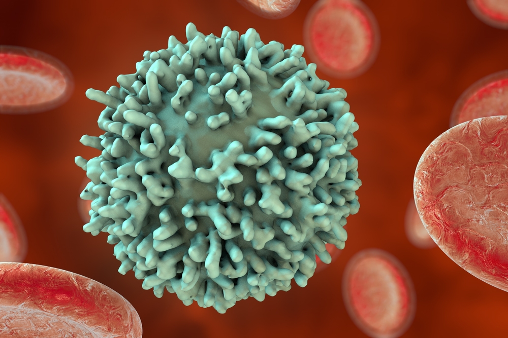
A significant difference between asymptomatic SCNI and non-SCNI individuals were found in a subset of immune cells known as CD4+ effector T-cells. These cells, also called T helper cells, play a critical role in regulating the immune response.
The similarity in cells isolated from SCNI patients was particularly pronounced with proinflammatory cells, such as those called Th17 and Th1 cells.
To validate these findings, the researchers isolated and analyzed cells from a second independent group of 115 early MS patients who had not been treated for the disease. Results showed some of the same changes observed in the twin studies, including the more pronounced populations of Th17 and Th1 cells.
Thus, the findings point to an essential role of subsets of T-cells in triggering MS disease.
“Taking these data together, by controlling for the dominant influence of genetic and local environmental factors, and by focusing on [early-stage] MS, we were able to reveal peripheral MS-related immune signatures pointing to a pivotal role of effector CD4+ T cells during disease initiation,” the researchers wrote.
“Insight into the immunological mechanisms associated with the initiation of the disease is relevant not only to the therapy but also for prevention of the disease,” they added.

Leave a comment
Fill in the required fields to post. Your email address will not be published.