
New 3D Model May Help in Promoting Myelin, Preventing Its Loss
Written by |
A new 3D model of the human nervous system is meant to mimic key processes in the development of the myelin sheath — the fatty coating around nerve cells that is damaged in multiple sclerosis (MS) — to help with research into treatments that promote myelination.
This model might also be useful in preventing demyelination, or the loss the protective myelin sheath, its researchers noted.
Investigators described their model in the study, “iPSC-derived myelinoids to study myelin biology of humans,” published in Developmental Cell.
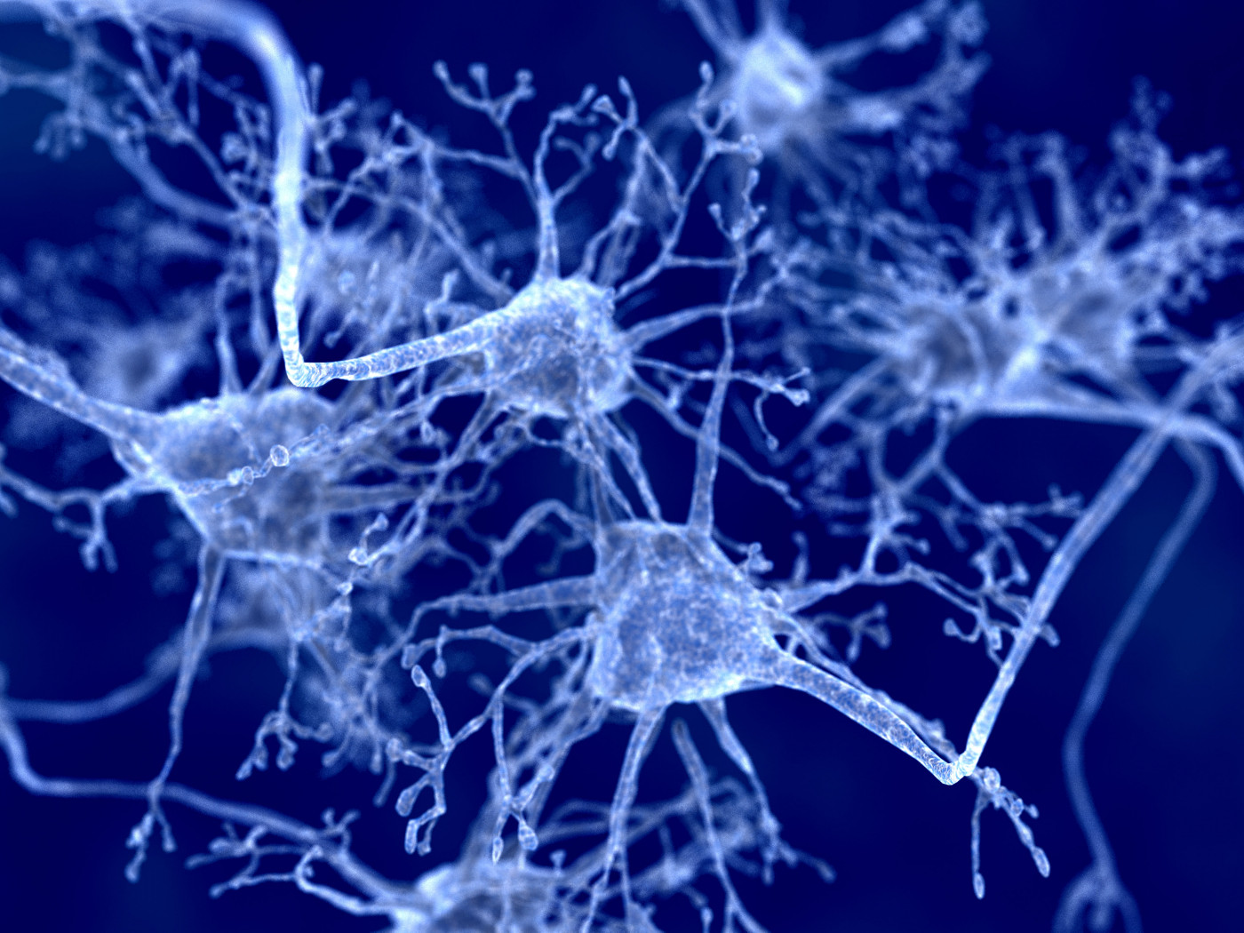
Nerve cells, or neurons, work in the nervous system to send the electrical signals that ultimately control thoughts and movements. These cells have long projections, called axons, that are coated with myelin, an insulating sheath that helps the cell more efficiently send signals.
The myelin sheath, as such, is critical for proper neuronal function, and in MS is damaged by erroneous inflammatory attacks. But models that capture processes in myelin’s development and its activity within the body are limited.
As the name suggests, an “organoid” is a laboratory model that is meant to resemble a little organ. Different kinds of cells are grown together in a three-dimensional structure that mimics how the cells would naturally be organized in an organ.
Induced pluripotent stem cells (iPSCs) — which can be engineered to grow and differentiate into other cell types — were used to create the organoids. iPSCs are a specific kind of stem cell created by researchers in a lab from other types of cells, usually skin cells.
By giving iPSCs a specific collection of biochemical cues, the researchers created organoids meant to resemble tissue in the spinal cord. Notably, these organoids included both neurons and oligodendrocytes, the cells that make the myelin sheath.
The researchers termed their model “myelinoids,” short for “myelinating organoids.”
Through a battery of experiments and imaging tests, they showed that active myelination was occurring in their models. In other words, they demonstrated that the oligodendrocytes were producing myelin to cover the neurons, a process known as myelination.
Myelination in these models also responded as expected to treatment with a toxin known to affect myelin production, leading to demyelination.
“We aimed to establish an induced pluripotent stem cell (iPSC)-derived, spinal cord-patterned model of myelin formation that enabled investigation of myelin development, disease, pharmacological interventions, and interrogation of activity-regulated myelination in a human context,” the team wrote.
In further experiments, the researchers characterized the models’ architecture, and how myelination changed over time. They also created a version of the model using iPSCs derived from a patient with a genetic disorder that affects myelination, and showed that the model recapitulated disease-specific changes.
“These data show that iPSC myelinoids can be used to model disorders of [myelination],” the team wrote.
Researchers hope to use these myelinoids to study diseases like MS, with the ultimate goal of finding treatments that address myelin formation.
“Now we have the capability of studying human myelination experimentally, a major goal is to identify drugs that can promote myelination. We believe that this new approach could be a huge boost to the toolbox that allows us to do this effectively,” Owen Gwydion James, PhD, a study co-author with the University of Edinburgh, said in a press release.
While this model is a step forward, some limitations were noted. First, because the model is meant to mimic spinal cord biology, it may not be well-suited for studying myelination in other locations, like certain brain regions.
Second, like any laboratory model, the myelinoids lack the full complexity of the human body. Particularly, their models do not contain a kind of supportive nervous system cell called microglia, the researchers wrote.
Finding ways to add microglia to the system, they wrote, “would provide an opportunity to study the role of microglia in myelin development, degradation, and repair, the role of which remains largely unknown.”


Leave a comment
Fill in the required fields to post. Your email address will not be published.