Spinal cord lesions tied to higher risk of clinical relapse in MS: Study
Findings support routine use of spinal cord MRI for monitoring patients

The presence of spinal cord lesions — alone or with brain lesions — on MRI scans was associated with a higher risk of clinical relapse for multiple sclerosis (MS) patients over those with just brain lesions, according to a recent study. Spinal and brain lesions together were also predictive of disability progression.
The findings support the routine use of spinal cord MRI for monitoring patients and making therapeutic choices, the researchers said.
“Monitoring MS with spinal cord MRI may allow a more accurate risk stratification and individual treatment optimization,” they wrote in “The added value of spinal cord lesions to disability accrual in multiple sclerosis” which was published in the Journal of Neurology.
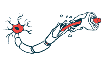
In MS, the immune system mistakenly attacks myelin, the fatty substance that surrounds and protects nerve cells, in the brain and spinal cord, together called the central nervous system (CNS).
As such, lesions, or areas of tissue damage with myelin loss, are found in various parts of the CNS. They can be visualized by MRI imaging to help diagnose and monitor the neurodegenerative disease.
Spinal cord lesions are often symptomatic and associated with neurological impairments, the scientists said. For this reason, spinal cord damage has been found to predict disability accumulation or the transition to progressive MS.
Spinal cord lesions and risk of relapse
There are technical challenges associated with acquiring and interpreting spinal MRI images, however. Generally speaking, a spinal cord MRI is not recommended for routinely monitoring MS, according to the researchers, who decided to explore its potential value with the disease.
They retrospectively examined MRI and clinical data from patients in the MS Registry of the Multiple Sclerosis Center at St. Andrea Hospital, Rome, where spinal cord MRI scans are regularly performed alongside brain MRI as part of follow-up.
Of 830 patients in the analysis, the most common disease course was relapsing-remitting MS (87.8%) and about two-thirds (67.3%) were on disease-modifying therapies (DMTs).
A total of 5,701 MRI scans were reviewed, with a median of six per patient over a median follow-up of seven years.
Most showed no evidence of active/inflammatory lesions, new lesions — those not present on a previous scan — or T2 lesions, a measure of total lesion load, in the brain or spinal cord. That’s likely because most patients were using DMTs, the scientists said.
Still, 17.9% of the scans showed T2 lesions exclusively in the brain, whereas 6% showed T2 lesions in the spinal cord, but not the brain, and 7.5% had them in both areas.
Active inflammatory lesions were found in the brain only for 11.5% of the scans, only the spinal cord for 5%, and both in 4.2%.
About a third of patients had at least one scan showing new T2 lesions in the spinal cord, and 26.1% had a scan with new inflammatory lesions. By comparison, 62.5% had a scan with new T2 lesions in the brain and 46.6% had new inflammatory lesions.
Asymptomatic inflammatory lesions, or those not associated with overt clinical symptoms, were more frequent in the brain alone (9.5%) than in the spinal cord alone (2.8%) or both (2%).
“The opportunity for a therapeutic reevaluation in patients with asymptomatic, isolated spinal [inflammatory] lesions would have been missed limiting the MRI protocol to brain monitoring alone,” the scientists said.
Ultimately, the presence of inflammatory lesions in the spinal cord alone or in both the brain and spinal cord were associated with an increased risk of relapse compared to active lesions in the brain only.
Also, 341 patients had disability accumulation after a median follow-up of 1.5 years. Disability worsening was associated with the presence of new T2 lesions in both the brain and spinal cord compared with such lesions in the brain alone.
“Overall, our data suggest that a routine MRI protocol, including both brain and spinal cord MRI, may help to detect a proportion of patients who would otherwise be considered stable,” the researchers wrote, adding such patients may need treatment plan changes, “with relevant implications on long-term disability outcomes.”


