FDA clears upgraded software to aid MRI analysis in MS
Neurophet software uses AI to measure disease-related changes
Written by |

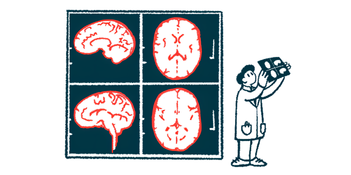
The U.S. Food and Drug Administration (FDA) has cleared an extended version of Neurophet Aqua, an MRI analysis software that uses artificial intelligence (AI) to measure disease-related changes in brain scans.
Earlier clearance enabled Neurophet‘s software to analyze brain atrophy using T1-weighed MRI scans in people with neurodegenerative conditions. These scans are often used with a contrast agent called gadolinium to identify active, inflammatory lesions in people with multiple sclerosis (MS), but they can also be used without gadolinium to quantify structural changes.
With the latest clearance, the software will include the additional features for analysis of MS and white matter hyperintensities, lesions that show up as bright, white spots on a type of MRI scan called T2-weighted fluid attenuated inversion recovery (FLAIR).
The aim is to help doctors quickly and accurately count and measure brain lesions caused by MS. The software also tracks changes in lesion number and size over time, with reports delivered within five minutes, according to the company.
“Neurophet AQUA’s advanced MS analysis technology significantly enhances efficiency and convenience for healthcare professionals, making it an indispensable tool in both diagnostic and prognostic stages,” Jake Junkil Been, Neurophet’s co-CEO, said in a company press release.
MRI analysis challenges
MS causes inflammation and damage in the brain and spinal cord, leading to symptoms such as fatigue and movement difficulties. Damage is visible as lesions on MRI, the gold standard for diagnosing and tracking MS progression.
“MS is a neurological disorder that heavily relies on MRI for both diagnosis and disease monitoring, and the McDonald criteria, the diagnostic criteria for MS, specifically includes MRI confirmation of lesions disseminated in space and time,” Been said.
There are many kinds of lesions in MS, and different types of MRIs may be needed to detect and quantify them. For example, T1-weighted MRI can be used to detect inflammatory lesions, as well as regions where permanent damage has occurred, which can show up as dark spots. T2-weighted scans are generally used to image the overall lesion load, including both old (inactive) and new (active) lesions.
MRI scans can also be used to monitor the loss of brain volume, which is a normal process that occurs as people age but is accelerated in people with MS due to ongoing damage and degeneration of nerve cells.
Subtle patterns and abnormalities in MRI scans can sometimes be missed, even by experienced radiologists and neurologists. Subjective interpretation and human fatigue may also cause variability in diagnostic outcomes, which can lead to delayed or missed diagnoses in some cases.
The incorporation of AI-based tools in the analysis of MRI scans can help overcome these challenges. By automating tasks such as lesion detection and monitoring and providing easy-to-read reports to healthcare professionals, these tools aim to save time and allow doctors to detect subtle changes that may require treatment adjustments.
“Neurophet AQUA’s advanced MS analysis technology significantly enhances efficiency and convenience for healthcare professionals, making it an indispensable tool in both diagnostic and prognostic stages,” Been said.
