
Brain sugar levels act as signal for myelin growth, study finds
Studies in mice, cells show link between glucose and oligodendrocytes
Written by |
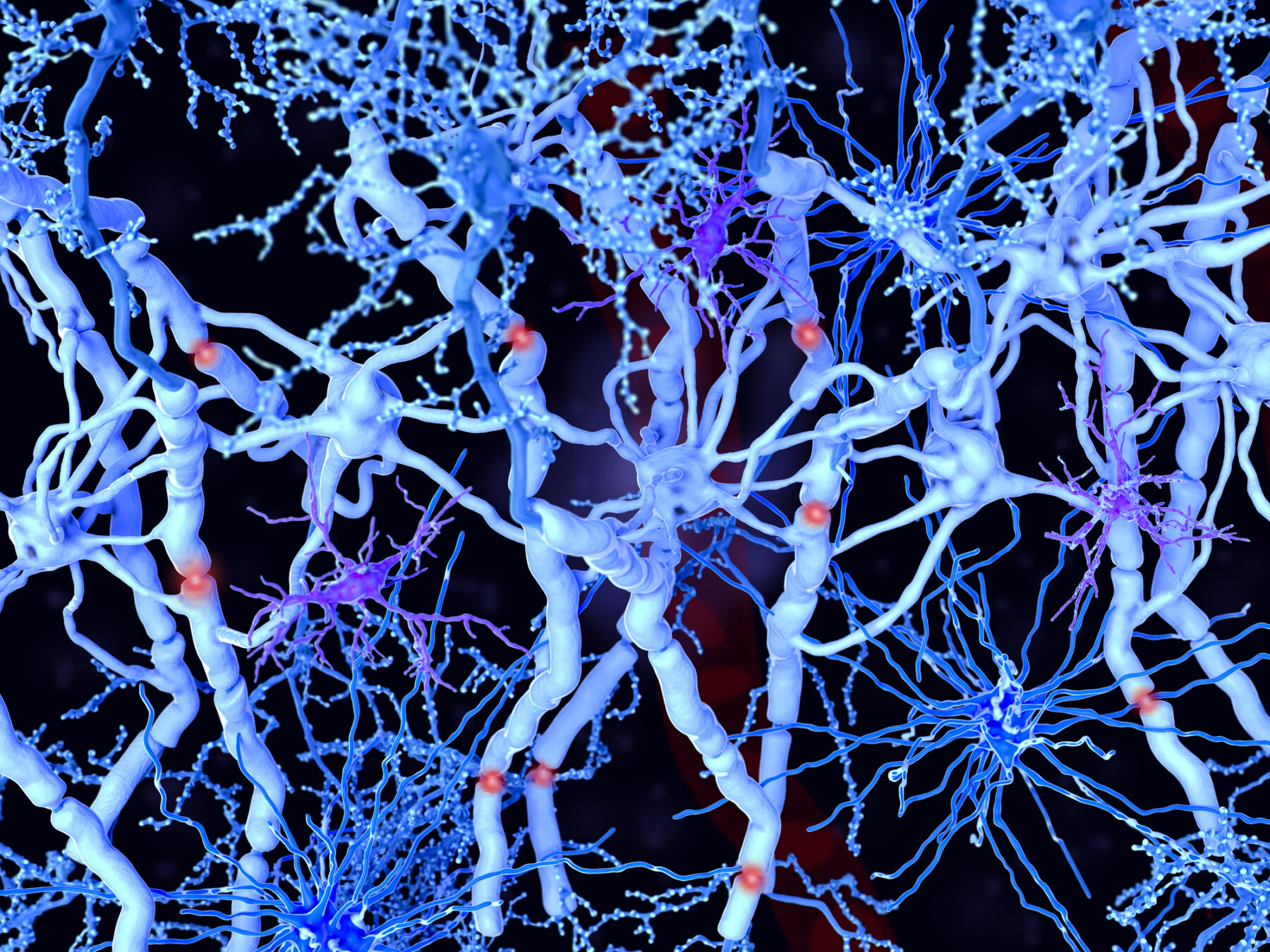
White matter in the brain is made up of myelinated axons, while oligodendrocytes form the myelin sheaths around the axons.
- Brain sugar levels signal oligodendrocyte progenitor cells to divide or mature for myelin production.
- OPCs can use ketone bodies from fat breakdown to produce myelin if glucose is unavailable.
- Ketogenic diets restored myelin production in mice, suggesting new metabolic strategies for MS.
The amount of sugar in the brain plays a key role in governing the activity of oligodendrocytes, the brain cells responsible for making the protective myelin coating around nerve fibers, according to a study done in mice and cell models. The findings could have important implications for understanding diseases such as multiple sclerosis (MS), which is characterized by inflammation that damages myelin in the brain and spinal cord.
Researchers found that molecules derived from sugar are essential for the growth of immature cells that can develop into oligodendrocytes. When these cells can’t rely on sugar, they can make functional myelin using alternative metabolic sources, notably ketone bodies produced when the body burns fat.
In fact, myelin production was decreased in a genetically engineered mouse model in which oligodendrocytes could not use sugar, but these deficits were restored by feeding the mice a ketogenic diet — a high-fat, low-sugar diet designed to get the body to burn fat instead of sugar for energy.
“Our findings show that glucose is not just fuel for the brain, it’s also a signal for the cells to divide,” Sami Sauma, PhD, co-author of the study and postdoctoral research associate at the City University of New York (CUNY) Advanced Science Research Center, said in a university news story. “We found that when glucose levels are high in a particular brain region, progenitors use it to drive proliferation. As glucose levels shift, the same cells switch gears and begin maturing. It’s a beautifully coordinated metabolic system that helps shape brain development.”
The study, “Glucose-dependent spatial and temporal modulation of oligodendrocyte progenitor cell proliferation via ACLY-regulated histone acetylation,” was published in Nature Neuroscience.

Molecular mechanics
Myelin is a fatty substance that helps protect nerve fibers and helps them send efficient electrical signals. In MS, damage to myelin disrupts nerve signaling, ultimately leading to MS symptoms. Oligodendrocytes can help repair damaged myelin, but myelin repair mechanisms are usually defective in MS for reasons that aren’t fully understood.
Oligodendrocyte progenitor cells (OPCs) are immature cells that are able to grow into mature, myelin-making oligodendrocytes. During brain development, OPCs either divide to make more OPCs or mature into myelin-producing oligodendrocytes.
The team conducted a detailed series of experiments aiming to understand the molecular mechanisms that control whether OPCs divide or mature.
By analyzing developing mouse brains, the researchers found that OPC activity showed a striking correlation with glucose (sugar) levels. In brain regions with high glucose levels, OPCs were mostly dividing to make more OPCs. But in regions with lower glucose levels, the cells were more likely to be maturing. This suggested that the cells were responding to glucose levels to regulate their activity.
Cells can use glucose for energy, but they can also break this sugar down to make other molecules, including one called acetyl-CoA. Acetyl-CoA is an important ingredient used in myelin production, and it is also essential for histone acetylation, which is a biochemical process that cells use to increase the activity of certain genes.
The researchers found that OPCs rely on acetyl-CoA derived from glucose in order to perform histone acetylation that turns on the genes that let them divide to make more OPCs.
Building on this finding, the researchers engineered mice whose OPCs lacked the enzyme ATP-citrate lyase (ACLY), which is needed to convert glucose into acetyl-CoA. In these mice, OPCs couldn’t rely on glucose to grow, and as a result, oligodendrocyte counts were abnormally low, and myelin production was decreased in early development.
However, as the mice grew, myelin production increased to levels typically seen in healthy mice, which suggests the OPCs were still able to grow into functional oligodendrocytes that could make myelin. Because acetyl-CoA is needed to make myelin, and these cells were making myelin even when they couldn’t get acetyl-CoA by breaking down glucose, the cells must have been sourcing this molecule from elsewhere.
In further tests, the researchers found that OPCs used acetyl-CoA generated by the breakdown of ketone bodies. Building on this finding, they fed a ketogenic diet to mice lacking the ACLY enzyme, which helped normalize myelin production during early development.
“Although OPCs depend on [acetyl-CoA derived from glucose] for histone acetylation and proliferation, [oligodendrocytes] are less discriminatory as to the source of [acetyl-CoA needed for] myelin synthesis,” the scientists wrote.
The researchers hope this new understanding of the biochemistry that controls myelin-making cells will pave the way toward better understanding and treatments for diseases like MS.
“This study reveals that the same cell lineage interprets different metabolic signals at distinct stages of development,” said Patrizia Casaccia, MD, PhD, study co-author and director of the neuroscience initiative at the CUNY research center. “By understanding how glucose and alternative energy sources regulate proliferation and myelin formation, we are uncovering new metabolic strategies that could be harnessed to protect myelin in the developing brain and even promote repair in disease states.”


Mark Collins
My body used to heal back after an attack, but not anymore. Wish it would start again.
Rana Imran
Hi dear
I'm from Pakistan
My son is a neuro patient
Can you guide me about my son
Pauline Lofkin
When my hubby, a Type 1 Diabetic, was first diagnosed with Diabetic Neuropathy around 2009 at the Royal Exeter and Devon hospital, Exeter, UK, the consultant said that he would have to have a a biopsy taken from his spine in order for him to determine if my hubby was suffering from DN or MS as they are very similar. The diagnosis of DN was made and my husband was faced with years of decreasing health issues. We had a neighbour who suffered from MS too so learned some bits from her about MS . Reading this article is interesting. I can see something in there that perhaps comes from the years of attending appointments with my hubby. I hope that you will be able to transfer research to other conditions that are similar to MS like Diabetic Neuropathy and begin to obliterate any conditions like MS to improve lives for people. My husband was in so much pain, he would cry. In the end he found it hard to give up smoking. He said it helped relieve the pain. He died in 2024, under so much morphine, I'm sure it helped him pass.
Steve Hards
Clearly this has potentially interesting implications for people with MS but unfortunately it's pretty incomprehensible to a non-specialist. I therefore asked AI to explain its implications and this was the summary: "These findings suggest that metabolic strategies, such as the ketogenic diet, could potentially be harnessed to promote repair in disease states like Multiple Sclerosis (MS). However, it is important to note that these specific results were observed in mice and cell models, and researchers are still working to understand how these metabolic signals can be safely used to protect and repair myelin in humans". Is that about it?
James P Devine
Would the ketogenic also help peripheral neuropathy?
Mabahlakoana Makuta
Does the research on MS indicate that it was wrong for medical professionals to insist that diets that include sugar and fat is bad for our health. Were we given wrong information about sugar and fat?
Edwin
This makes much sense.Since the brain is mostly fat then we should eat lots of good fats and less carbs
Stella Walker-Sharland
I cannot but wonder why sugars help brain cells grow/mature in mice, why does that not appear to be the case in humans. I was told that high blood sugar levels were a key player in dementia and related brain problems. I am wondering if the sugar that crosses the blood brain barrier has a different molecular structure.
Monica Rodriguez
Does this finding help people with PPMS as well? My son has the worst form of MS contracted it at 23.
Marian Corkish
I am very interested in any news about the progress in research about multiple sclerosis. My daughter was diagnosed with it 3 years ago, aged 40
Rex Bobi
A phenomenal finding that will have enormous benefit, achieved through great and determination of tireless scientists like Patricia Casaccia and many others. Congrats. 👏🏻👏🏻👏🏻👏🏻👏🏻
Terrie J Fachetti
I was told my myelin, sheath and axioms were destroyed by chemo Would it work for me ?Because I have no axioms?
John Dozois
Can you please make the findings more understandable to the non scientists please?
Sugar and ketosis and the myelin sheath and glial cells
Is regulating glucose levels through fasting good for people with MS?
Michael Pass
So, are they promoting a keto diet, or saying a high-sugar diet is helpful? Or, if you are on a keto diet, your body will just adapt? Is there a clear benifit to high-sugar diet versus keto? Is this about adaptation of getting Acetyl-CoA through different biochemistry? I may need to reread, but it's not clear in the summary portion.