Real-world Data of Gilenya Treatment Validates Slowed Brain Shrinkage as Disease Progression Measure
Written by |
A real-world study of Gilenya (fingolimod) in relapsing multiple sclerosis (MS) confirms benefits of the treatment seen in clinical trials. The Novartis-sponsored study also demonstrated that measures of brain shrinkage can be used in a clinical setting to evaluate disease progression.
The data, presented at the American Academy of Neurology (AAN) 2017 Annual Meeting in Boston, showed that Gilenya effectively prevented several measures of disease activity and progression for more than a year.
The Phase 4 study, called MS-MRIUS (Multiple Sclerosis and clinical outcome and MRI in the U.S.), included 590 patients treated with Gilenya in 33 U.S. clinics. Patients were followed for a median of 16 months. During this time, 59.6% of 586 assessed patients achieved NEDA-3 status — a term that implies no relapses, no new or enlarged brain lesions, and no disability progression.
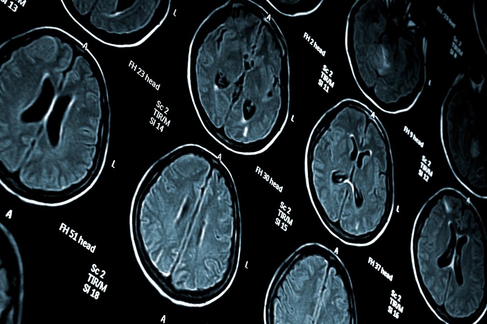
In addition, 325 patients were assessed for NEDA-4, which also included magnetic resonance imaging (MRI) measures of brain shrinkage. In this group, 37.5% achieved NEDA-4.
Breaking up the components of this composite measure showed that among those with NEDA-4, treated with Gilenya, 86.5% had no relapses, 91.1% had no disability progression, 79.7% had no new or enlarged brain lesions, and 58.2% had a brain shrinkage rate similar to that seen in people without MS.
“These data build on the wealth of clinical and real-world evidence that show Gilenya is a highly efficacious, long-term treatment option for controlling disease activity in relapsing MS,” Vas Narasimhan, Global Head of Drug Development and chief medical officer at Novartis, said in a press release.
The study also validated, for the first time, a method for brain shrinkage assessment that is available at neurological clinics.
“Measuring brain shrinkage has historically been dependent on specialist brain scanning techniques,” Narasimhan said. “These groundbreaking new data showing brain shrinkage can be reliably measured by routine MRI scans that have the potential to change how this key measure of disease progression is monitored, to ultimately help patients and physicians observe and manage treatment success and outcomes.”
The study design and an interim data analysis were published in December 2016, and included information on 252 patients.
Researchers now suggest that monitoring of brain shrinkage could be adopted in daily clinical practice.