
#EAN2018 – Both Brain and Spinal MRIs Needed to Monitor MS Inflammation, Study Says
Written by |

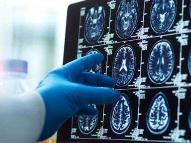
Magnetic resonance imaging (MRI) used to assess inflammation in multiple sclerosis (MS) patients should include scans of the spinal cord and not be restricted to the brain, because brain scans alone risk underestimating disease progression, a study suggests.
These results were shared in the presentation, “Measuring disease activity in Multiple Sclerosis: do we need spinal cord MRI?” (abstract on page 71) given at the 4th Congress of the European Academy of Neurology (EAN) that recently concluded in Lisbon.
When MS is suspected in a person, guidelines recommend an MRI of the spinal cord and brain to reach a diagnosis. But to monitor disease progression in people with confirmed MS, only brain MRIs are typically taken.
Whether spinal MRIs are also effective at monitoring progression is still controversial.
The researchers hypothesized that “using brain MRI only might fail in detecting inflammation in a proportion of patients.”
They investigated the frequency of spinal cord acute inflammation, and whether this occurred independently from brain activity. Their study looked at data on MS patients who underwent at least two different MRIs (brain and spinal cord) at least 30 days apart, and had MRI reports available.
Join our FORUMS to discuss the latest research and trials!
Inflammatory activity was defined by the presence of at least one gadolinium-enhancing lesion according to its location (either in the brain, the spinal cord, or both). Gadolinium (Gd), also called a “contrast” agent, is a harmless chemical compound given by injection that passes from a patient’s bloodstream to sites of active inflammation in the brain or spinal cord where it lights up, creating a visual image on an MRI that flags an inflammation site.
The team analyzed 5,717 scans, of which 4,537 (79.3%) showed no Gd enhancement.
Out of the remaining 1,180 scans, 651 (55.2%) showed brains with only Gd-enhanced lesions, and 232 (19.7%) had Gd-enhanced lesions in both the brain and spinal cord.
However, 297 (25.2%) of these scans showed Gd-enhanced lesions only in the spinal cord.
Disease-induced spinal cord damage can occur independently of damage taking place in the brain, the study concluded.
“Our study demonstrates that inflammatory activity can be detected frequently in SC [spinal cord] and occurs in approximately 25% alone. Limiting MRI monitoring to brain, underestimates inflammatory activity,” the researchers wrote.
“MRI monitoring of SC [spinal cord] in clinical practice will allow neurologists to switch treatment to more powerful drugs” as needed, the team added.


Dave Uherek
When you say spinal chord, do you mean cervical, thoracic and lower lumbar areas ( defining the entire spine) ? Should the MRI be taken with and without gadolinium enhancement? Thanks for any answers.
Karri
Hi Dave,
As an MS veteran, I can say with a high degree of certainty that the C-spine is what should be monitored. I had my annual MRI’s with both brain and C-spine, gad and no gad, studies done since 2001. My neuro has stopped doing annual MRI’s after gadolinium was found in cadaver brains (she now does MRI on an as-needed basis).
I hope this info helps.
Garden lady
Gadolinium is hardly harmless. Researchers have found it deposited in brain tissue, and health issues can result. Talk with your Neuro!
Erika Faber
While reading this article, I was unsettled reading the following: "gadolinium (Gd), also called a “contrast” agent, is a harmless chemical compound given by injection that passes from a patient’s bloodstream. . ."
Within the last few years, there has been talk that the contrast that is used, "Gd", is actually harmful and can remain in your system for x amount of time. I don't have the names or quotes, but when I heard this, it made me question getting any MRI, CT, etc., with the contrast Gd.
So, following the progression of MS is important, however, in many cases, I think that we with the disease are in tune with our bodies and know when the disease is getting worse. I have had MS for at least 26 yrs and know when it is progressing. I am having increased cognitive impairment, decreased mobility, and a list of other symptoms that were not present 5 or 10 years ago. Other conditions were eliminated. Also, some symptoms have increased in intensity. So, to take a risk of getting an MRI ~ brain and spine, seems like a waste of time and money as well as taking a risk that I would rather not take. My health is not optimal so why take a chance at adding to declining issues. So, to say that Gd is a harmful chemical. . .please research this drug and double check complaints about this contrast before putting it out there that it is harmful when it is not. Thank you.
Here is a page I encourage you to check out!
FDA Panel Backs New Warning for Gadolinium Contrast Agents
Manufacturers could be required to conduct new studies
SAVESAVED
https://www.medpagetoday.com/radiology/diagnosticradiology/67811 for the full story:
Dt
Gadolinium being a harmless chemical compound is not true.
Doctors recommend it because of the convenience to do. if you care about gadolinium build up in your system only authorize it if new lesions are detected.
Pat Rowbotham
Why is time and money being wasted on "research", to "discover" what your average 16 year old biology student could tell you. Lesions on the brain versus myelin depletion caused by inflammation to the spinal chord. Which will put you in a wheelchair? For 25 years I have repeatedly told various neurologists exactly where the inflammation in my spine was, but was never given an MRI scan. As I now cannot walk , I don't qualify for any drugs that may save my arms. Isn't that discrimination against the disabled? I would pay for the drugs but nobody will write me a prescription. The whole system is sick!
Susanne E Rettell
Wow! Just wow! I was dx'd with Ms in 1999 but I am sure I had it since 1981. Lately, things have really gone downhill. Cognitive wise and other issues. My legs and hips are always in pain. When I tell them they just say its because of the bad discs in my back. While that may be partially true I do believe a lot of it is the MS. Last week I was woke out of a dead sleep at 4 in the morning with the worst cramps in my ankles, calves, and feet. I was in screaming pain. I quickly chewed a valium up as I was desperate for it to stop. It seemed like it lasted forever. The next day it felt like someone kicked the shit out of my legs they were so dang sore it was unbelievable. The second day I was unable to walk. What a helpless feeling that was as I have no help here. All they want to do is give you more and more drugs. I was supposed to get the Acthar Gel but it was denied by my insurance company. I am also a cancer survivor and regular steroids could make any cancer cells grow rapidly if they were in my system. Seriously your just their guinea pig and they don't care if they kill you. I hate this! Every day I pray they find a cure so everyone who suffers from this can have their life back. MS sucks and so do all their drugs.
Karen
I too, suffer from that terrible pain in my legs, calves and feet. I’ve had times where I’ve had to put ice packs on the outer parts of my thighs and calves. I am also declining both cognitively and mobility wise. I’ve had MS for 25 years. I was diagnosed at 30.
This is such an evil disease that not only slowly takes away our abilities, but also sucks away at our souls.
We are guinea pigs and at their mercy.
itasara
I also have had more feet and ankle cramps and some calf cramps when I wake up especially in the morning. I take b12, and calcium, and magnesium and other supplements. I have read,howeer that a lot of people have cramping especially as one ages, so it may not be an MS problem. Then again it might. I just don't know. Could be that I don't do a lot of exercise... my bad!
Alan
I was diagnosed on March 18, 2013, the first couple of years it was an MRI every 3 months and then it was 2x’s a yr. I switched to a Dr I thought specialized in MS. My last appt he declaredI did not have Optic Neuritis. I went to my optician and he said I did. I went to an Eye Dr and after doing the exact same test said ?I did not have it, only dry eye. Meanwhile 90% of the time my eyes blur or I have little things float across my eye.
Back to my Neuro, I use a cane outside the house because my legs are weak and my balance comes and goes. I also have a broken back. He had me walk down the hall telling me to straighten up. My hips need to be replaced as well so I do hunch over at times. But the one little hall walk he goes back in and using a little microphone he dictated to the computer that all of my leg problems are he believes to be from ”other issues” not MS. I broke my back in 1985. I can tell you what living with chronic pain is like without insurance. I was given all kinds of pain meds but I hated the side effects and they never did a thing for my pain levels. After awhile you learn that self medicating with booze or pot help more than anything because if it makes you brain dead you don’t think about the pain. I know that my back never made my feet feel like they are frozen in a block of ice or my legs feel as though the bones are being crushed. It didn’t help that neither doctors gave me any education about the disease. There is a small group here in Palm Springs ACT for MS that has lots of information a library and bingo. I love the fact that it exists but I also suffer with ADHD, I have all my life. I barely graduated high school. But now I am supposed to educate myself about a disease. I just this week figured out what counts as a relapse, kind of. The first time I had extremely bad leg pain for over 24, I did not know about the 24hr deal but I was given an MRI and was told that I could either just suffer through it or I could go sit in the hospital overnight and get iv steroids! “If it would make me, meaning my mind feel better”. I was made to feel like a fool and was really wasting peoples time. So each visit when asked if I’ve had any relapses I have always said no. Mostly because I thought a real relapse had better be bad enough to be hospitalized or
I had to be as sick as I was when I first got sick and was in the hospital for 3 months. Now with the murders in control of the Congress and White House I am terrified I am going to lose my Disability, my insurance, my home, my life. I just did the Briova survey. I wish that Soc Security took into account all that that survey did. Not just the physical but the mental. Damn ?I can’t remember my meds or which day of the week it is half the time. My last review was a Phychiatrist, lmao I actually spelled that right. I used to be a champ at spelling now I can’t remember words much less hiw to spell them. But I’m only RRMS and no new lesions since the last MRI whenever that was lol.
Oh Susanne I know how you feel and because it is MS that we have I also don’t know how you feel. That is the f’d up thing about this disease is that it can be so extremely different with each person but we are treated the same. The government doesn’t care that this disease may allow lots of us to continue working while lots of us can’t and the others may not be able to tomorrow. I’ve dealt with major depression my whole crappy life. Now add Cog Fog, which the Dr never asks about, along with the extreme emotional ups and downs, mood swings that make PMS a cake walk and anxiety from hell. But even though those are also thought and considered to be MS related no Neuro asks about. We are stuffed with pills that only make our Drs and Big Pharma richer. I laugh when I read shit that hospitals or other Health websites talk about your Healthcare “Team”. Non of my Dr’s talk to the others, they won’t do it. I just lost my pain dr that took me ages to trust due to the fact that Anthem Blue Cross is screwing them so I can either go out of network which I can’t afford or go back to one .i didn’t like. My last complaint is that I see you were diagnosed in 1999, I’m like you I am positive I was having symptoms years before. But I see how long you’ve dealt with it and while you give me hope I am also so damn tired of it all that I can’t imagine living like this for that many years. Waiting and wondering when or if the next one will hit, or how bad will it be. Or just living with this pain much longer. I have the worlds best husband. He tries so freaking hard to take care of and provide for us. He indulges me hoping that I will be happy. In turn he’s gotten to wipe my butt, clean my diaper when I had no control over either bowels. He has drained my cath bag after working a 12 hr shift. I once thought that it would be AIDS that would first torture and then kill me but the MS is winning that battle. I have what should be a perfect life but I can’t. I can’t remember not being in pain so bad I’wish a truck would run me over because that would stop the pain. If it wasn’t for my hubs I’d be out on the 10 thumbing a ride to Neverland. I don’t pray anymore, after a slow realization of who he is and what he has become I had an awakening and my god died recently. So now I wish and use affirmations, so I wish you many days of being pain free. I also wish you thousands of those little moments that make life worth living. I wish a cure for this God Awful disease. But most of all I wish you peaceful heart soul and mind. Be well!
Love Alan
itasara
I had two MRIs one in 2005 when I was diagnosed of spine and brain, and one in 2007 of the brain. Went to neurologist for past 12 years - not a specialist, who did not feel I needed any more MRIs since I was doing so well. Then this year he said get one. The hospital said they do not use Gadolinium any more but a different variety that does not have similar side effects. The results were hard to read-- different machines than 12 years ago - but he suggested I go to MS specialist. I plan now to continue with the specialist. I should have done so in the first place. I am still waiting to hear if she wants to change anything - I've been on Copaxone since day one. If there were any significant changes, because I have no record in the past 12 years there are no historical markers to see when changes took place and if therapy should have been changed.