
Cerebrospinal Fluid Neurofilament Predicts CIS and RRMS Progression, Study Contends
Written by |

Cerebrospinal fluid levels of neurofilament light chain, a protein associated with nerve cell damage, can predict disease progression in people with clinical isolated syndrome (CIS) and relapsing-remitting multiple sclerosis (RRMS), a Swedish study found.
Higher levels of neurofilament light chain (NFL) in the fluid that surrounds the brain and spinal cord, the cerebrospinal fluid (CSF), were associated with the appearance of new brain lesions on magnetic resonance imaging (MRI) scans, and also loss of brain volume during a four-year follow-up period.
The study “Neurofilament levels, disease activity and brain volume during follow-up in multiple sclerosis” was published in the Journal of Neuroinflammation.
Better prognostic biomarkers, ones that are able to predict MS disease activity and identify who is at higher risk of developing new lesions or flare-ups, are needed.
Prior studies indicated that brain volume loss detected with MRI, and markers present in the CSF or blood, may be used as tools to assess disease progression and activity.
One of these potential biomarkers is NFL, a protein exclusive of nerve cells and one of the major byproducts released to the CSF and blood during nerve cell death. That makes NLF a potential “universal biomarker” of neurodegeneration.
CSF levels of NFL correlate with long-term prognosis, and are a risk factor for conversion from CIS to relapsing MS. But blood tests are preferable because they are less invasive compared to CSF sampling and MRI scans. In fact, increasing evidence suggests that measuring NFL levels in the blood can be a good test for prognosis and disease monitoring of MS.
Based on these promising findings, researchers now examined the correlation between NFL levels in the serum (blood) and CSF. The team also looked for links to other neurodegenerative and neuroinflammatory biomarkers, disease activity measured on MRI scans, disability level, and relapses.
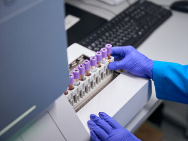
A group of 41 patients with newly diagnosed CIS or RRMS, referred to the department of Neurology, University Hospital of Linköping, Sweden, were monitored for a four-year period. Analyses were compared to a group of 22 healthy people used as controls.
Patients underwent clinical examination, blood and CSF sampling, as well as MRI exams at baseline and at one, two, and four years of follow-up. Immunomodulatory treatment was maintained throughout the study.
Serum and CSF levels of NFL correlated significantly in patients at baseline; both were significantly higher in patients when compared to healthy participants.
However, NFL levels in the CSF were stronger predictors of disease activity than in the serum. CSF levels were more sensitive in identifying patients with “no evidence of disease activity-3” (NEDA-3) — a definition of absence of active disease — NEDA-3 is a defined as no relapses, no brain MRI activity, and no confirmed disability worsening.
CSF levels of NFL predicted active disease with a sensitivity of 95% at one year, 93% at two years, and 81% at four years. In contrast, sensitivity of serum levels was lower: 76% at one year, 74% at two years, and 72% at four years.
Also, NLF levels seem to predict which patients with CIS will progress to RRMS. Levels of NFL and two other biomarkers, CXCL10 and MMP-9, present in CSF were significantly higher in patients who converted to RRMS during follow-up, compared with non-converters. Contrarily, NFL and other markers in the serum did not differ.
Other biomarkers in CSF, such as CHI3L1, CXCL1, CXCL10, CXCL13, CCL22, and MMP-9, also correlated with disease activity either with loss of brain volume or the appearance of new brain lesions.
“CSF-NFL predicts and reflects disease activity better than [serum]-NFL,” the researchers wrote. “Our findings further add to the accumulating evidence that CSF-NFL is a useful biomarker in CIS and RRMS and should be considered in the expanding NEDA concept,” they added.
Other biomarkers that need further study include CSF CXCL10 and CHI3L1 for prediction of disease activity and brain volume loss, respectively, researchers noted.


Leave a comment
Fill in the required fields to post. Your email address will not be published.