Harnessing T-cell Subtype May Suppress Immune Responses in MS, Study Suggests
Written by |
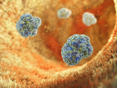
Though known mainly for killing tumor and virally infected cells, a T-cell subtype may restrain immune responses and be protective in autoimmune diseases such as multiple sclerosis (MS), according to new research.
The study, “Opposing T cell responses in experimental autoimmune encephalomyelitis,” was published recently in the journal Nature.
The involvement of different subtypes of immune T-cells has been shown in MS patients and in the experimental autoimmune encephalomyelitis (EAE) mouse model of the disease. A coordinated T-cell response to gluten also was reported in patients with celiac disease, but whether that response occurs in MS is undetermined.
Researchers from Stanford University School of Medicine addressed this gap by using the EAE model, in which animals are injected with a protein called myelin oligodendrocyte glycoprotein (MOG) to trigger MS-like disease.
Within 10 days, the blood levels of T-cells containing the CD4 marker — and specific for a MOG peptide (MOG35–55) — peaked, and later declined to baseline.
“When T-cells encounter a pathogen, or antigen, single cells that recognize some part of the pathogen divide and produce many copies of themselves,” Naresha Saligrama, PhD, the study’s lead author, said in a press release. “This suggested that a specific population of cells were responding.”
But a different subset, containing the CD8 marker, also showed an initial increase, followed by a drop to pre-EAE amounts. Importantly, these CD8+ T-cells suppressed the proliferation of CD4+ T-cells.
“We absolutely think that something like this is happening in human autoimmune diseases,” Mark Davis, PhD, the study’s senior author, said. “It represents a mechanism that nobody’s really appreciated. There’s this whole subset of CD8 T-cells that has a suppressive function.”
The investigators then addressed which peptides triggered the CD8+ T-cell response. Because initial tests with 350 myelin peptides were not successful, the team expanded the pool of candidates to roughly 5 billion with a molecular approach called yeast display, which generates peptides attached to individual cells.
“We are crowdsourcing the T-cells. We’re asking the T-cells, as the disease is progressing, what they are interested in,” Davis said.
This led to the identification of two peptides — HDR and SMRP — that were recognized by these suppressive T-cells. Injection of the peptides before, after, or alongside MOG resulted in significantly lessened disease severity, or even no disease. Experiments in a different model, the experimental autoimmune uveitis, did not show similar benefits, indicating that the eased manifestations are specific to EAE, the researchers noted.
Subsequent analysis then revealed that the suppressive properties of CD8+ T-cells were associated with an increased expression of regulatory genes, and with the expression of surface proteins linked with reduced immune function.
Supporting the findings in mice, analysis in cells isolated from MS patients also showed a marked boost in blood levels of suppressive T-cells and, to a lesser extent, of inflammatory CD4+ T-cells.
In summary, the “results suggest that the induction of autoreactive CD4+ T-cells triggers an opposing mobilization of regulatory CD8+ T-cells” in mice, and probably in humans, the researchers wrote.
“There’s this whole subset of CD8 T-cells that has a suppressive function. If we could mobilize those cells to function more effectively in patients with autoimmunity, then we’d have a novel treatment for diseases like multiple sclerosis,” Davis said.
The team has plans to assess if these suppressor CD8 T-cells also are involved in other autoimmune diseases, including celiac disease.