
Hypoxia-Measuring Technology in MS Could Be Potential Biomarker for Treatment Responses
Written by |
In a new study entitled “Reduced cortical microvascular oxygenation in multiple sclerosis: a blinded, case-controlled study using a novel quantitative near-infrared spectroscopy method,” a team of researchers at the Hotchkiss Brain Institute, Cumming School of Medicine, University of Calgary investigated whether frequency domain near-infrared spectroscopy technology can measure the potential lack of oxygen (hypoxia) in brain grey matter regions of multiple sclerosis (MS) patients. The study was published in the journal Scientific Reports.
Hypoxia (a term describing the insufficient concentration of oxygen in the blood and tissues) has been implicated in several diseases, including Alzheimer’s disease and MS. Its role in brain disease pathogenesis is potentially linked to the hypoxia modulatory action of inflammation responses in the brain, a key step in MS (MS is an autoimmune disease that affects the brain and spinal cord; the disease is triggered by an inflammatory and immune attack to the myelin sheath, a protective layer that surrounds nerve cells). However, the study of hypoxia in the brain has been halted due to technological constrains.
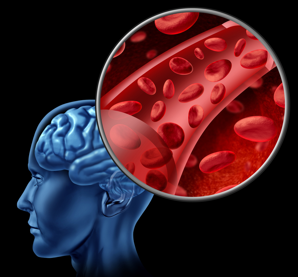
Frequency domain near-infrared spectroscopy (fdNIRS) is a system that quantifies microvascular hemoglobin saturation (StO2), a measurement that may be used as a potential marker of hypoxia in MS (98% of oxygen is transported in the blood mainly reversibly bound to hemoglobin, measuring hemoglobin saturation in brain small vessels indicates oxygen delivery efficiency and microvascular-associated performances).
In this study, a team of researchers determined if reduced StO2 values were associated with the pathophysiology of MS. To this end, the team measured fdNIRS in a cohort of 72 MS patients (including relapsing remitting (RRMS), secondary progressive, and primary progressive MS patients) and controls , focusing specifically in the brains’ grey matter.
The researchers detected that StO2 is reduced (more than 2 times) in SPMS and high disability RRMS patients, suggesting a relationship between StO2 and clinical disability in MS patients. These data also suggest that there may be hypoxic regions in an MS brain.
As a result of these findings, researchers demonstrated that fdNIRS technology is useful in the study of MS and other neurological disorders, and highlighted that measuring StO2 is a potential early response biomarker to assess treatment response in MS patients.


Leave a comment
Fill in the required fields to post. Your email address will not be published.