
MS Lesions in Cerebellum Ably Predict Disability Levels and Disease Progression, Study Suggests
Written by |
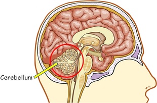
The cerebellum, found in the back of the cerebral cortex, is like a "mini-brain" for coordination and smooth movement.
Damage to the cerebellum in people with multiple sclerosis (MS) is due more to the death of actual nerve cells than the destruction of white matter connections, a new study out of Italy suggests. The article, which challenges previous ideas about how brain damage in MS occurs, is titled “MRI-detectable cortical lesions in the cerebellum and their clinical relevance in multiple sclerosis“ and appeared in Multiple Sclerosis Journal.
MS is characterized by lesions on white matter, in what known as the loss of myelin. This means that the insulating substance that wraps around neurons is damaged, impairing communication within the nervous system and causing symptoms such as loss of movement, coordination and vision, tingling sensations, and even cognitive and emotional problems. White matter lesions have been a focus of MS research and treatment, but increasing evidence points to cells being lost in MS as well. In the cerebellum, this cell loss is known as cortical lesions.
The cerebellum, found in the back of the cerebral cortex, is like a “mini-brain” for coordination and smooth movement. Although neurologists tend to focus on MS lesions in the cerebral cortex, lesions in the cerebellum may also be important. However, these lesions can be hard to detect, due to the cerebellum’s physical location.
Scientists led by Alice Favaretto, of the Department of Neurosciences DNS, The Multiple Sclerosis Centre–Veneto Region, University Hospital of Padova, Italy, studied a total of 40 people with relapsing-remitting MS (RRMS). The team found that cortical lesions (the kind in which damaged neurons are found) were more common in the cerebellum than white matter lesions. In addition, cortical cerebellum lesions could be used to predict the level of disability in people with MS.
Cortical lesions (CL) “are detected by PSIR [phase-sensitive inversion recovery] in the cerebellum of the majority of MS patients, are more than WML [white matter lesions], increase with disease progression and strongly correlate with the cerebellar EDSS [expanded disability status scale]. Thus, the observation of CL in the cerebellum of MS at clinical onset might be useful for prognostic and therapeutic aims,” the scientists wrote.
This research indicated that cerebellar cortical lesions are clinically relevant, and may be important for neurologists to monitor in evaluating MS disease progression and disability.


Spiro spyratos
Terrific. For two decades we've been told it's a demyelination disease when clearly, axonal loss has been the hallmark. We need to focus on it being a neurodegenerative disease and finding ways of regenerating neurons, i.e. Stem cells, neural progenitor cells, etc. also, finding ways to reduce degeneration with antioxidants targeting mitochondrial loss. Refocused research
Anastasios Chortis
Very well said - Focus on remyelination, Mesenchymal stem cell-derived neural progenitors (MSC-NPs) and ways that reboot and repair the immune system not suppress the immune system.
Brian Melton
Well said Spiro
Tracey
Very well said. What their Focus has been on has not worked or had any resolution and no proof of help. Focus more on our own bodies to fix it I. E Stem Cells
Jeff
And what about radical diets and exercise like Wahls Protocol to juice up our mighty mitochondria? Or do studies show stem cells regenerate myelin more? Please, more stories about studies like all of the above, thank you.
Amit
It also shows lack of understanding of the complexity of this disease, despite centuries of it's prevalence.
Marcie
Yes, maybe now those of us who don't show active lesions/increase in lesions on our MRI even though we're clearly experiencing a deterioration in functional ability will be taken seriously by our doctors instead of being called alarmists and told we're NOT getting worse. I'm the one living in this body after all; I know when I can no longer hold onto things without dropping them or falling and staggering like a drunken sailor!
Jeff
That's crazy. Deterioration for people with progressive MS is often NOT accompanied by new lesions. Axonal loss does not correspond with lesions. Call the MS society and/or talk to another doctor, you deserve better health care.
Grace
We can only hope.
Cindy Marlene Morales
Ugh... i'm with you on that! So tired of hearing, but your MRIs are stable. Then explain to me why i don't feel stable!
Hector
Hello from Texas,
Very well written article here.
Susan Van Zandt
I am told I have PPMS and have had active lesions on my last 2 MRI's (18 months apart). Recent in left cerebellum.I have no impairment on my left side at all, only weakness and foot drop in my right leg. No measureable progression in 18 months noticeable by me. More a frustration when my leg gets tired and I am not ready to quit what I am doing!! If it wasn't for the lesions and lack of other another diagnosis, I wouldn't believe it was MS at all.