Gray Matter Lesions Affect Cognition in Japanese MS Patients as Well, Study Says
Written by |

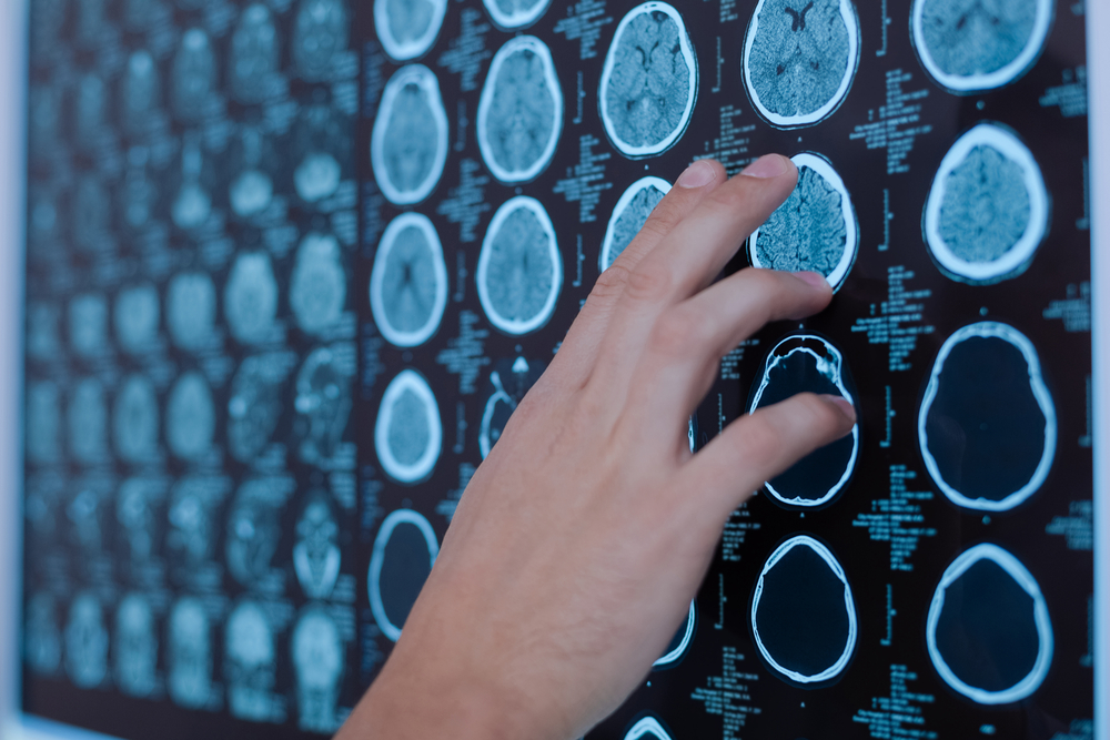
People in Japan with lesions in the cerebral cortex due to multiple sclerosis (MS) were found to have greater cognitive problems, or difficulties thinking, than those without lesions in this area of mostly gray matter that surrounds the brain, a study reports.
Lesions confined to the cortex contributed more to verbal and visual memory problems. Lesions that also extended into white matter — the brain area typically affected by MS — were linked to difficulties with attention, information processing, and working and visual memory as well.
These findings mirror similar results reported in patients of European ancestry.
The study, “Contribution of cortical lesions to cognitive impairment in Japanese patients with multiple sclerosis,” was published in the journal Nature Scientific Reports.
MS is caused by immune system attacks on the myelin sheath, the fatty layer that surrounds nerve cells and helps in the transmission of nerve impulses. This damage shows up as spots called lesions on magnetic resonance imaging (MRI) scans in areas of the brain known as the white matter — so-called due to the presence of myelinated nerve cells.
Lesions can also be found in the surrounding grey matter area, known as the cerebral cortex. These so-called cortical lesions have two subtypes: those within the cortical (cortex) area only, called intracortical lesions (ICLs), and those that involve both the cortex and white matter, called leukocortical lesions (LCLs).
Cortical lesions have been linked to cognitive difficulties in people with MS. Cortial lesions are found in higher numbers and greater volumes in relapsing-remitting MS (RRMS) patients with cognitive issues compared to those without. The presence of LCLs are more strongly associated with cognition problems than ICLs.
Much of this research has focused on people of European descent, with few studies investigating cortical lesions and cognitive impairment in those from an Asian background.
Scientists based at Kyushu University in Japan investigated how the different types of cortical lesions affected cognition in Japanese people with MS.
A total of 61 patients (mean age at disease onset, 29.4) were included in their study, of which 42 were diagnosed with RRMS and 19 with secondary progressive MS (SPMS). All were clinically stable without a relapse for at least one month before MRI scans.
In addition to the MRI scans, the cognitive abilities of patients were assessed using the Brief Repeatable Battery of Neuropsychological Tests (BRB-N), which is a series of tests of verbal learning memory, visual-spatial learning and memory, attention, working memory, information processing, and verbal abilities.
The Expanded Disability Status Scale (EDSS) assessed each patient’s disability level. Through questionnaires, signs of apathy, fatigue, anxiety and depression were also reviewed. An overall measure of a person’s cognitive abilities was calculated as a cognitive impairment index (CII) score.
Results showed that in most of the BRB-N cognitive tests, as well as in the overall CII score, MS patients with cortical lesions scored significantly poorer compared to those without these lesions.
Patients with ICLs had significantly lower scores on the visual-spatial learning and memory test, the auditory test, and in the verbal abilities test, and a higher overall CII score than those without gray matter lesions.
Likewise, patients with LCLs — compared to those without gray and white matter lesions — had higher CII scores, and worse scores on visual-spatial learning and memory, attention, information processing, and auditory tests, as well as on the verbal learning memory test.
Overall, those with LCLs showed greater cognitive impairment compared to patients with ICLs.
“Leukocortical lesions were more extensively associated with cognitive dysfunction in various domains than intracortical lesions,” the researchers wrote.
A statistical analysis identified the number of cortical lesions and EDSS scores as the primary factors linked to an overall CII score of cognitive difficulty.
No differences were found in the apathy scores, fatigue measures, or anxiety and depression scales between MS patients with and without cortical lesions.
“Our results demonstrate that the presence and number of CLs [cortical lesions] are robustly associated with cognitive dysfunction in Asian MS patients,” the researchers wrote.
“LCLs were more significantly associated with the impairment of cognitive domains preferentially affected by MS, including attention, information processing, and working and visual memory, while ICLs may contribute more to verbal and visual memory impairments,” they added.
Based on the results, the team concluded that the “presence and number of CLs could be relevant risk factors for cognitive decline in Japanese MS patients.”


