
Boosting Cholesterol Recycling in Brain Facilitates Myelin Repair, Study Says
Written by |
New research in mice suggests that poor recycling of cholesterol in the brain impairs the repair of myelin, the protective coat surrounding nerve cells that is lost in multiple sclerosis (MS).
Pharmacological stimulation of cholesterol synthesis by brain immune cells — called microglia — boosted the regeneration of myelin, supporting its potential as a new therapeutic strategy for MS.
The study, “Microglia facilitate repair of demyelinated lesions via post-squalene sterol synthesis,” was published in the journal Nature Neuroscience.
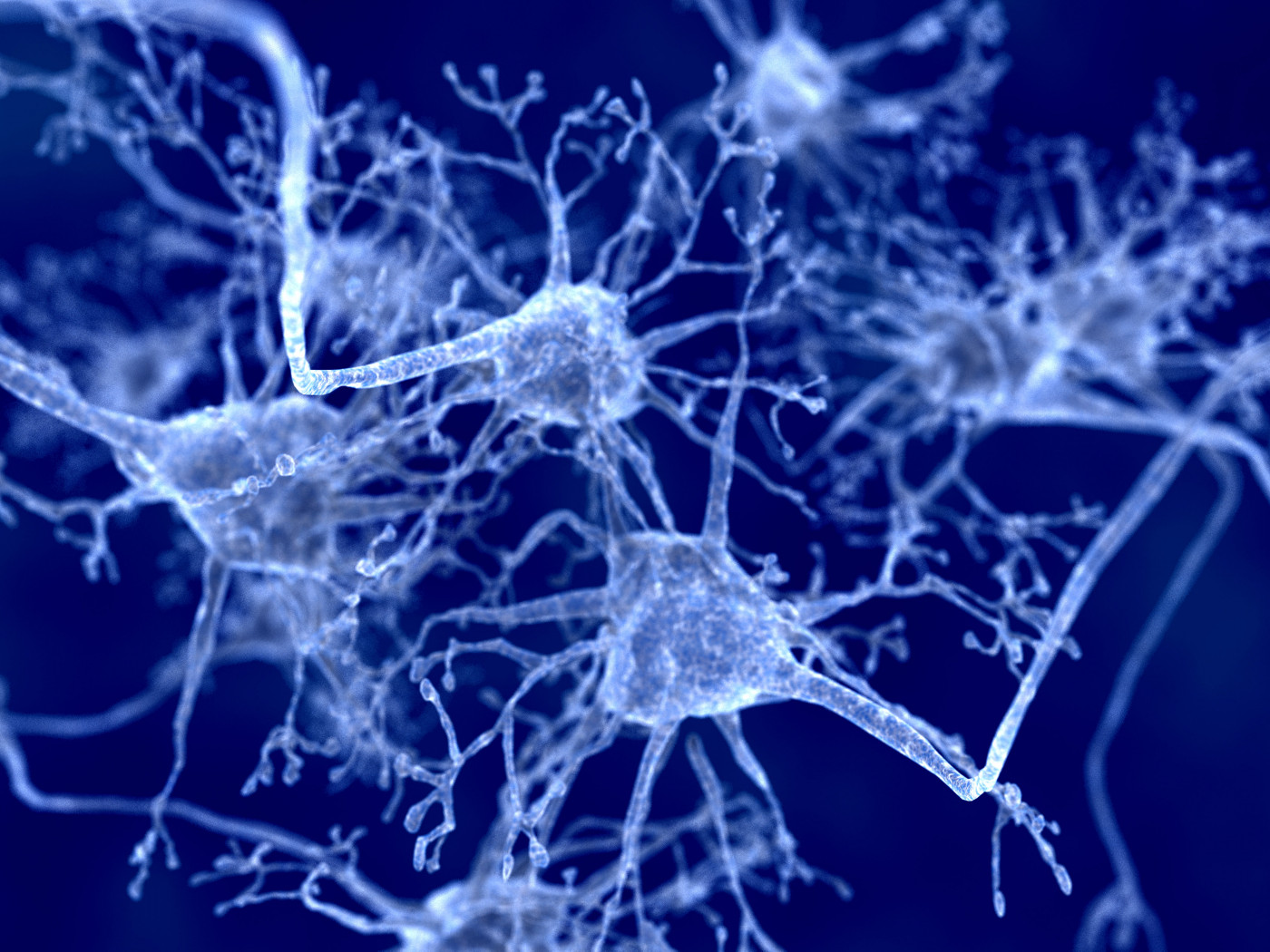
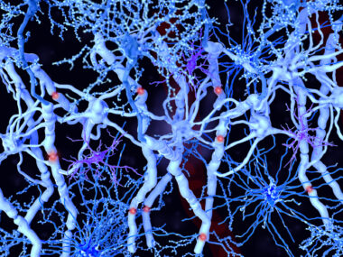
MS is characterized by the loss of myelin, a cholesterol-rich sheath that protects nerve cells. Damaged or defective myelin in the central nervous system or CNS, comprised of the brain and spinal cord, release cholesterol-rich myelin debris that is taken up and recycled by microglia. These immune cells of the brain use that debris for the production of new myelin, which is mediated by myelin-producing cells called oligodendrocytes.
“This efficient repair mechanism probably takes place all the time in healthy individuals,” Gesine Saher, at the Max Planck Institute for Experimental Medicine in Göttingen, Germany, and the study’s lead author, said in an institute press release.
Here, the researchers investigated whether the production of sterols, like cholesterol, in different cell types of the CNS play a role in the repair of demyelinated lesions.
The team analyzed the corpus callosum, the area that connects the two sides of the brain, in cuprizone-treated mice (an animal model of MS) at two stages of demyelination — both acute and chronic — and remyelination.
The analysis of cholesterol-related genes revealed that cholesterol synthesis in microglia cells was required for remyelination after acute, or early, demyelination.
Elimination of “cholesterol synthesis in CNS microglia … resulted in severely impaired remyelination following acute demyelination,” the researchers wrote.
Next, the researchers deleted genes linked with the synthesis of cholesterol in microglia cells and macrophages — scavenger cells of the immune system. Of note, microglia and macrophages are collectively called phagocytes.
The cells were still able to absorb cholesterol from damaged myelin but were unable to recycle it, the scientists found. The phagocyte cells were locked in a pro-inflammatory state.
“We wanted to find out why phagocytes stop making the absorbed cholesterol available again,” said Stefan Berghoff, the study’s first author.
The team then investigated whether sterol production in microglia cells played a role in resolving inflammation in MS, using the cuprizone and the experimental autoimmune encephalomyelitis (EAE) model, which is another mouse model that mimics MS inflammation in humans.
The findings showed that the levels of pro-inflammatory genes were increased, while anti-inflammatory and genes involved in cholesterol recycling were reduced.
These genes were all involved in the LXR signaling, a key pathway involved in the resolution of inflammation. That suggested that a defective LXR signaling could underlie the ongoing inflammation in MS.
Further experiments showed that in response to demyelination, microglia cells increase the production of an intermediate product of cholesterol synthesis, called desmosterol, which induces the activation of the LXR signaling.
This, in turn, leads to an equilibrium of cholesterol synthesis and the return of microglia cells to an anti-inflammatory and a pro-regenerative state supporting remyelination by oligondendrocytes, the myelin-producing cells.
“As a signalling molecule, this precursor molecule [desmosterol] not only caused the mobilization of cholesterol for the formation of new myelin sheaths but also helped establishing a pro-regenerative environment,” Berghoff said.
Data from cells isolated from human patients with MS showed signs of activation of the LXR signaling, supporting the findings in mice.
The researchers then tested the effects of pharmacological stimulation of cholesterol synthesis in myelin restoration. In lab tests, adding an early intermediate of cholesterol synthesis — squalene — to cells collected from the spinal cord boosted the production of myelin by oligodendrocytes.
Importantly, administering squalene to EAE mice increased the presence of anti-inflammatory markers and boosted the repair of demyelinated lesions. This occurred in parallel with an increase in the activity of genes linked with cholesterol synthesis, suggesting new therapeutic strategies for myelin repair in MS.
“These in vitro and in vivo data support the concept that squalene supplementation ameliorates inflammatory demyelination by activating LXR signaling to resolve inflammation and enhance repair,” the researchers wrote.
Moreover, a combination of squalene with interferon beta-1b (IFN beta-1b), a standard first-line therapeutic option for MS, ameliorated disease scores in EAE mice better than each treatment alone. A triple treatment with the combination of squalene, IFN beta-1b, and a potent LXR activator led to a more pronounced lessening of EAE clinical disability.
Overall, “our results in mice revealed the terpene squalene as a new potential factor in the therapy of myelin diseases such as multiple sclerosis,” Berghoff said.
Saher said the findings may be helpful for people with MS.
“Our results suggest that this treatment has a similar positive effect in humans as in mice,” Saher said.
Of note, previous studies suggested that a Mediterranean diet may reduce disease severity in MS. Researchers noted that this diet — rich in the consumption of fish, fruit, and vegetables — can lead to a squalene consumption of 200 to 400 milligrams per day.
“Thus, squalene supplementation should be further investigated as a therapeutic strategy in human MS,” the team added.


Leave a comment
Fill in the required fields to post. Your email address will not be published.