New Spinal Cord Lesions Can Be Evident in Stable MS Patients
Written by |

Asymptomatic damage to spinal nerves occurs even in clinically stable cases of multiple sclerosis (MS) and carries an increased risk for further lesions, a recent study suggests.
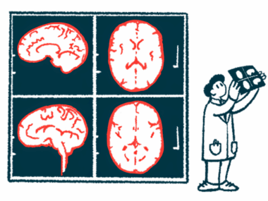
Although a firm link between the asymptomatic loss of myelin in the spine and worsening disability remains to be found, this work suggests that monitoring clinically stable MS patients via MRI scans may prove useful.
The study, “Recurrence and Prognostic Value of Asymptomatic Spinal Cord Lesions in Multiple Sclerosis,” was published in the Journal of Clinical Medicine.
The loss of myelin — a protective cover over nerves that helps them conduct electric impulses — characterizes MS. Areas of myelin loss are called lesions, and increase the risk of disability when they occur in important areas with tightly packed nerve fibers like the spinal cord.
Research is starting to show, however, that some spinal lesions appear without new or worsening MS symptoms — the so-called asymptomatic spinal lesions, or a-SL. This raises the question of whether these lesions can serve as markers for MS activity and treatment response, and therefore whether to include a spinal MRI in routine patient monitoring.
Researchers in Italy and Switzerland analyzed the medical records of relapsing and progressive MS patients to assess the frequency, recurrence, and prognostic value of a-SL.
The team identified 300 people in the MS patient registry at the Multiple Sclerosis Center in Lugano, Switzerland, who had undergone an initial spinal MRI (T1) followed by another nine to 36 months later (T2), and were clinically stable between T1 and T2, with stability defined as an absence of relapses and disability progression measured by the expanded disability status scale (EDSS). All patients had a clinical follow-up (T3) at least six months after T2.
Of these 300 patients, 249 had relapsing-remitting MS (RRMS), 36 had secondary progressive MS (SPMS), and 15 had primary progressive MS (PPMS).
Asymptomatic spinal lesions were found in 45 patients (15%) at T2 over a median of 14.3 months, and occurred in both relapsing and progressive cases. Of these 45 people, six (13.3%) had higher EDSS scores — indicating greater disability — at T3, as did 53 of the 255 patients without a new a-SL at T2 (20.8%).
Relapses were relatively infrequent between T2 and T3, occurring in five of 38 RRMS participants with an a-SL at T2 (13.2%) and in 21 of the remaining 211 RRMS patients without an a-SL (9.9%).
A statistical analysis indicated that a-SL presence did not associate with a greater risk of relapse or increased disability.
The risk of new spinal lesions or a new brain lesion, in contrast, did correlate with the presence of an a-SL between T1 and T2.
Of 266 patients who underwent a brain MRI at T3, 69 (25.94%) showed new brain lesions. New spinal lesions appeared in 27 of 242 patients (11.16%) with spinal MRI findings at T2 and T3. Overall, brain lesions were about 1.63 times more frequent, and spinal lesions seven times more frequent, in people with evidence of asymptomatic lesions at T2 than those without.
Asymptomatic brain lesions (a-BL) occurring between T1 and T2 also correlated with higher risk of developing new brain lesions.
“The prognostic value of a-SL predominantly concerned the future accumulation of spinal rather than brain lesions,” the investigators wrote, “and similarly, a-BL better predicted the occurrence of brain lesions.”
Although spinal involvement tends to associate with disability progression, this study did not find a link between disability progression and asymptomatic lesions.
“In conclusion, we found that asymptomatic spinal lesions are detectable in approximately 15% of clinically stable MS patients over a median period of 14 months. If present, a-SL confers an increased risk of future accumulation of brain and spinal demyelinating lesions, particularly at the spinal level, that may contribute to disability accrual over the long term,” the researchers wrote.
“A link between asymptomatic spinal demyelination and disability worsening remains to be established, as well as whether the appearance of a-SL should prompt changes in disease modifying treatments,” the team added.
Of note, a previous study found that an accumulation of spinal lesions between one and three years after first evidence of MS correlated with higher EDSS scores 15 years later, suggesting that a longer follow-up might be needed for a relationship between asymptomatic lesions and disability outcomes to be evident.
“Despite this,” the researchers concluded, “our data suggest it might be useful to consider spinal MRI in the monitoring of clinically stable MS patients, particularly in case of spinal involvement.”