
Brain Volume, NfL Level Changes May Predict Disability in Relapsing MS
Written by |

A model that considers changes in brain volume and serum neurofilament (sNfL) levels during the early stages of multiple sclerosis may help clinicians to determine an individual’s likely progression with relapsing forms of MS, a study suggests.
“We were able to build reliable, robust models capable of accurate predictions of future clinical worsening over time in individual patients, while taking respective baseline values into account for each subject,” the researchers wrote, stressing that their work also showed no single biomarker could predict a range of future outcomes.
The study, “CNS Atrophy Predicts Future Dynamics of Disability Progression in a Real-World Multiple Sclerosis Cohort,” was published in The European Journal of Neurology.
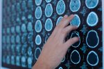
Clinicians currently base MS treatment plans on brain MRI scans that assess specific areas of inflammation within the brain. However, frequent MRI scans of broader brain regions in conjunction with monitoring alterations in brain volume have been shown to provide a clearer picture of disease severity and optimize treatment strategies.
Besides MRI scans, assessing blood levels of sNfL — proteins that maintain the structural integrity of nerve cells — may also help clinicians develop appropriate treatment plans, since past studies linked high blood sNfL levels with greater MS severity.
More individualized treatment is a key goal of MS management. But to effectively develop such treatment plans, clinicians must have a sense of how a patient’s disease will progress over time, and there are currently no models to reliably predict MS disability progression.
A team of researchers in Switzerland sought to determine whether a model that considered the extent of brain volume loss (atrophy) via regular MRI exams and measures of sNfL levels over initial visits could predict disease progression and severity over time in people with relapsing MS.
Data from a large group of people with relapsing-remitting MS (RRMS; 140 patients) and secondary progressive MS (SPMS; 43 patients ) being followed at the University Hospital in Basel were collected
Study participants’ mean age was 46.4, their disease duration was 15.7 years on average, and most (67.2%) were females.
All patients underwent annual functional tests, including the expanded disability status scale (EDSS), a measure of MS disability, the 9-hole peg test (checking hand and finger dexterity), and the timed 25-foot walk test, a measure of walking ability.
Over a minimum of two years (mean, 2.2 years), all underwent three MRI scans, and their sNfL levels were examined at the time of the third MRI. These MRIs gave an idea of brain volumes at the beginning of follow-up, and of subsequent atrophy (shrinkage) of different brain areas, including white matter, gray matter, the thalamus, pallidum, striatum, and cervical spinal cord.
Gray matter contains the cell bodies of neurons (nerve cells), whereas white matter houses myelinated axons, the projections neurons use to connect to each other. The thalamus is a region of the brain that controls sensation, while the pallidum regulates voluntary movements.
After the three MRI exams, patients were followed for up to 11 years, during which they had a mean of six visits.
After reviewing the data and developing various models, the researchers found that brain atrophy assessments could predict disease worsening over time.
Among patients with SPMS, faster decreases in cervical spinal cord volume and increases in lesion load were significantly associated with greater disability over time, defined as worsening EDSS scores.
No association between brain atrophy and EDSS changes over time was seen in people with RRMS, but lower thalamus volumes on the first MRI scan (baseline measure) significantly associated with poorer walking ability in this group.
Among the SPMS patients, higher lesion volume and lower pallidum volume at baseline, as well as faster progression of atrophy in grey matter, white matter, and the cervical spinal cord were all significantly associated with slower timed-25-foot walking test scores.
In terms of hand and finger dexterity, lower gray matter volume at baseline significantly associated with worse dexterity in the dominant hand in subsequent years for RRMS patients, while faster increases in lesion load were significantly associated with poorer dexterity in the non-dominant hand.
No such associations were found in people with SPMS for the dominant hand. For the non-dominant hand, higher striatum volumes and sNfL levels at baseline, faster atrophy progression in white matter and the spinal cord, and faster increases in lesion load were significantly associated with poorer future dexterity.
“An important conclusion drawn from our predictive models is that there is no single one-fits-all biomarker that predicts every future outcome. Depending on the clinical outcome of interest, different variables seemed to be crucial predictive factors,” the researchers wrote.
“Future research in MS patients should rather focus on global assessments of the CNS [central nervous system] implementing as many regions of interest rather than isolated atrophy metrics in order to increase clinical relevance of the findings,” they added.
A study limitation was the use of retrospective data. Some patients were also lost to the study during follow-up, which could have led to incomplete data sets and possible bias.
“This study demonstrates the capability of short-term MRI-metrics to accurately predict future dynamics of neurologic disability progression in a large real-world relapse-onset MS cohort,” the researchers concluded.
Leave a comment
Fill in the required fields to post. Your email address will not be published.