
AI tool effectively measures MS disease activity from 1 MRI scan
MindGlide to be used in research to assess brain damage, treatment
Written by |
A new artificial intelligence (AI) tool called MindGlide can accurately calculate, from a single MRI scan, multiple aspects of brain damage related to multiple sclerosis (MS) — even when employed with routine scans that are not typically used to monitor such damage.
The development and validation of the tool was described in a study titled “Enabling new insights from old scans by repurposing clinical MRI archives for multiple sclerosis research,” which was published in the journal Nature Communications.
“Using MindGlide will enable us to use existing brain images in hospital archives to better understand multiple sclerosis and how treatment affects the brain,” Philipp Goebl, PhD, first author of the study at University College London, said in a university press release. “We hope that the tool will unlock valuable information from millions of untapped brain images that were previously difficult or impossible to understand, immediately leading to valuable insights into multiple sclerosis for researchers.”
A further goal, according to Goebl, is to use MindGlide and other AI tools to help current MS patients.
“In the near future, [we hope] to better understand a patient’s condition through AI in the clinic,” Goebl said. “We hope this will be possible in the next five to 10 years.”

Patients typically need multiple scans to assess MS damage
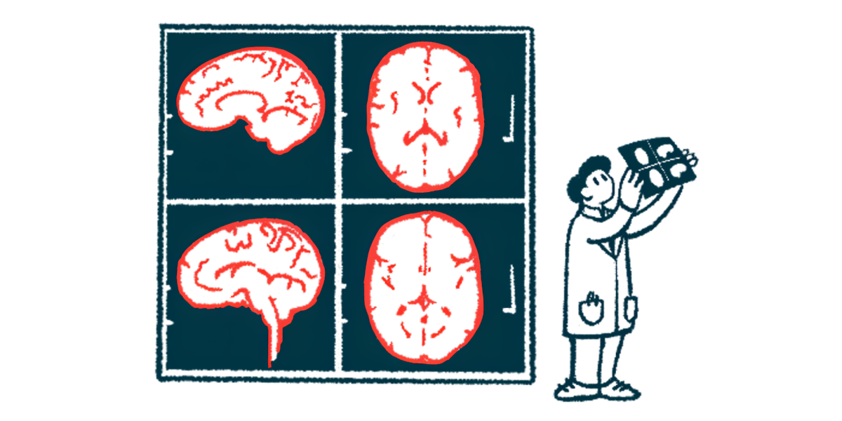
MS is caused by inflammation in the brain and spinal cord that damages healthy nervous system tissue. MRI scans are a key tool used to track MS disease progression and determine how a person with the neurodegenerative disease is responding to treatment.
On MRI scans, MS-related areas of brain damage are visible as white or dark spots, which are known as lesions. Traditionally, lesions have been identified by having experts manually examine MRI scans. This approach is time-consuming, however, and getting the best results usually requires experts to look at several different types of MRI scans of the same brain — meaning patients have to sit through multiple scans.
This need for repeated scans is not only inconvenient for patients, but also makes it more logistically difficult to run clinical trials tracking the effects of treatments on lesions.
MindGlide was developed with the aim of creating a tool that could accurately identify lesion load in the brain based on a single MRI scan — regardless of the type of scan. Having such a tool would make the process easier.
According to the researchers, “simplifying MRI analysis, particularly through single [MRI scans], can expand research opportunities from hospital archives, allowing analysis of previously acquired routine-care scans and potentially making clinical trials less costly.”
Like other AI tools, MindGlide was developed by first starting with a massive dataset — in this case, the researchers used more than 4,000 MRI scans. These scans were derived from nearly 3,000 people with various types of MS, and acquired with nearly 600 different MRI machines.
These data were fed into a computer alongside a set of mathematical tools, called algorithms, which the computer uses to identify patterns among the information it’s given. By identifying these patterns, the computer is basically able to learn how to identify lesions.

AI tool tested, validated using nearly 15,000 scans from 1,000 patients
The trained tool was then tested on nearly 15,000 scans from 1,000 patients followed in clinical trials or as part of routine care. The data demonstrated that MindGlide was better than other state of the art computer tools at identifying MS lesions in accordance with expert reviewers.
The AI tool also was effective at determining the effects of treatments in clinical trial data. In one analysis, the researchers compared MS lesion load and brain atrophy, or shrinkage, in people with primary progressive MS (PPMS) who were treated with either Ocrevus (ocrelizumab) or a placebo. This was also done using scans that have never been used for this purpose.
The results showed that patients given Ocrevus had significantly lower increases in lesion volume and slower rates of brain volume loss across different types of scans. The correlation between lesion load and disability levels was greater with MindGlide than with the standard of care AI tools.
We developed and validated [an MRI scan type]-agnostic deep learning model that can quantify MRI biomarkers from routine care and clinical trial datasets from varying single [MRI scans].
Other analyses used MindGlide to compare lesion rates in a trial of simvastatin against a placebo in people with secondary progressive MS (SPMS). Results using different scans consistently showed no difference in lesion volume changes over time, but a significant slowing in brain volume loss with simvastatin. Consistently, trial data showed that simvastatin failed to slow disability progression in SPMS.
“We developed and validated [an MRI scan type]-agnostic deep learning model that can quantify MRI biomarkers from routine care and clinical trial datasets from varying single [MRI scans],” the researchers concluded.
A notable limitation of the tool, the team noted, is that it only works on MRI scans of the brain — though MS also can cause damage in the spinal cord, which further contributes to disability. Collectively the brain and spinal cord are known as the central nervous system.
“Future work should expand our approach to the entire central nervous system,” the researchers wrote.
Leave a comment
Fill in the required fields to post. Your email address will not be published.