
Pregnancy hormone estriol promotes myelin repair in MS mice
Researchers call for clinical trials to explore potential treatment for remyelination
Written by |

Treatment with estriol, a hormone that’s produced during pregnancy, reduced disease severity and promoted myelin repair in the cortex — a key brain region affected in multiple sclerosis (MS) — in a mouse model of the disease.
Those are the main findings of the study, “Neuroprotection in cerebral cortex induced by the pregnancy hormone estriol,” published in Laboratory Investigation and conducted by a team of scientists at the University of California, Los Angeles (UCLA).
“This is the first study to identify a treatment that could repair myelin in the [brain’s] cortex, undoing some of the damage caused by MS,” UCLA stated in a university press release.

Based on these promising results, the scientists are calling for studies to examine the potential for this hormone as a treatment for women with MS.
“Estriol is an attractive therapeutic option,” the team wrote, noting that this hormone has been in use for decades as a therapy to help manage symptoms of menopause and has a generally good safety profile.
“Future clinical trials of estriol treatment as a neuroprotective treatment for MS are warranted based on preclinical and clinical data on efficacy as well as its safety track record, together representing a risk benefit ratio within the realm of other disease modifying treatments approved and in development for MS,” the researchers wrote.
Estriol is a form of estrogen that’s made by the developing fetus and placenta during pregnancy. Data from early clinical trials in women with MS have suggested that estriol has nerve-protecting properties, and it’s thought that increased levels of estriol may be one reason MS disease activity tends to lessen during pregnancy.
Now, the UCLA team used mice with experimental autoimmune encephalomyelitis (EAE) — a common laboratory model of MS — to investigate the effect of estriol treatment on disease progression and brain health.
Only female mice were tested
All of these experiments were done in female mice.
“We expect that estriol-treated male mice would exhibit similar effects to estriol-treated female mice,” the team wrote. But they chose not to conduct those tests because “as a potential therapy for MS, estriol treatment may lead to unwanted side-effects in male patients.”
The researchers found that starting estriol treatment shortly after disease onset resulted in significantly less-severe movement problems, compared with a placebo.
Estriol-treated mice also showed significantly less atrophy, or shrinkage, of the cortex, a brain region whose shrinkage in MS is linked to permanent disability worsening, than animals given a placebo.
The number of certain cortical neurons and the levels of PSD95 — a marker of healthy synapses (the connections between nerve cells) — were reduced significantly in EAE mice given a placebo relative to healthy mice. However, estriol treatment was associated with a normalization of these measures, reaching levels comparable to healthy animals.
“Together these findings demonstrate that estriol treatment after disease onset can restore neuronal and synaptic integrity in cerebral cortex,” the researchers wrote.
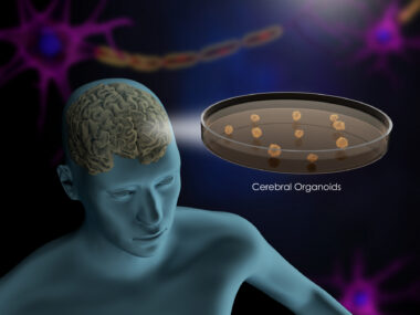
MS is characterized by the progressive damage and loss of the myelin sheath, the protective fatty layer around axons, or nerve fibers, that allows fast nerve cell communication. Data showed that mice treated with estriol had significantly less myelin damage in their brains than those given a placebo.
EAE mice given a placebo also showed obvious signs of axon damage in their spinal cords, while this damage was reduced significantly in mice treated with estriol.
More oligodendrocytes
Further tests revealed that estriol-treated mice had significantly lower activation of microglia, the brain’s resident immune cells that are thought to contribute to myelin loss, and significantly more oligodendrocytes, the cells chiefly responsible for making and repairing myelin.
In addition, the production of cholesterol, a fatty molecule that is a major component of myelin, by oligodendrocytes was increased significantly with estriol treatment.
“This demonstrated that treatment with [estriol] induced the expression of cholesterol synthesis proteins in oligodendrocytes, increased the number of remyelinating [myelin-repairing] oligodendrocytes, and restored myelin in the cerebral cortex,” the researchers wrote.
The team noted that prior trials that tested estriol in MS did not include measures of myelin repair. These preclinical findings “warrant investigation of estriol treatment to induce remyelination,” they wrote.
Estriol acts on cells by binding to a cellular receptor called estrogen receptor beta (often abbreviated ERB or ERbeta). The researchers showed that treating EAE mice with an ERbeta ligand, which activates the receptor, had similar effects to estriol treatment, including reduced disease severity, more myelin and oligodendrocytes, better synapse health, and less microglial activation.
“We found for the first time that both estriol and [ERbeta] ligand treatment reduced microglia activation in the cerebral cortex to not only induce remyelination, but also to protect synapses,” the researchers concluded. Based on these findings, they called for further trials to test the hormone as a potential therapy.


Leave a comment
Fill in the required fields to post. Your email address will not be published.