New Way of Growing Astrocytes from Stem Cells May Aid Research into Brain Disorders Like MS
Written by |

Researchers at The Salk Institute have developed a way to grow vital brain cells called astrocytes from stem cells, a potential breakthrough for basic and clinical research into several diseases, including multiple sclerosis (MS).
The study “Differentiation of Inflammation-responsive Astrocytes from Glial Progenitors Generated from Human Induced Pluripotent Stem Cells” was published in the journal Stem Cell Reports.
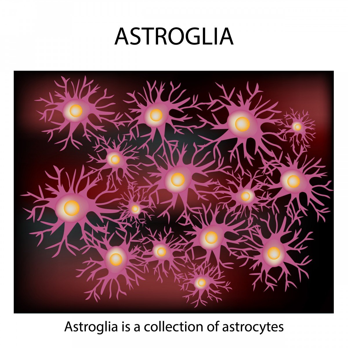
Research in neurosciences has been focused on neurons, the basic working unit of the brain. But neurons do not work alone, with a plethora of other cells carrying out vital functions in the brain. One is a group of star-shaped support cells called astrocytes.
Scientist working at The Salk Institute in California have now developed a method for generating astrocytes from stem cells.
“This work represents a big leap forward in our ability to model neurological disorders in a dish,” Rusty Gage, the study’s lead author and a Salk professor who holds the Vi and John Adler Chair for Research on Age-Related Neurodegenerative Disease, said in a press release. “Because inflammation is the common denominator in many brain disorders, better understanding astrocytes and their interactions with other cell types in the brain could provide important clues into what goes wrong in disease.”
Astrocytes provide neurons with energy and work as a platform to clean up their waste.They also have other functions within the brain, like regulating blood flow and inflammation.
Ways of differentiating astrocytes from human stem cells already exist, but are known to be laborious and functionally limited. Using this new method, researchers were able to generate astrocytes more efficiently — using the right “cocktail” of factors at the right time and settings. The functionality of these astrocytes is also closer to those in the brain, being, for example, sensitive to inflammation.
Astrocytes generated using this method are also able to be grown with neurons, which will allow researchers to study the interactions between both cell types to better determine what goes awry in disease.
“There are other methods for differentiating astrocytes, but our protocol arrives at inflammation-sensitive cells earlier, which makes modeling more efficient and straightforward,” said Carol Marchetto, a Salk senior staff scientist and study author.
Another important feature is that the cells — which will ultimately differentiate into astrocytes (called astrocyte precursor cells) — can be frozen. This allows researchers to keep a stock of cells that can be expanded later into astrocytes, without the necessity of going through the whole process of differentiation. In terms of laboratory experiments, the cells’ ability to be frozen will save researchers roughly six weeks of work.
The team performed tests to confirm the astrocytes’ behavior, and found that they functioned very much like astrocytes isolated from actual brain tissue. That is, they responded to neurotransmitters glutamate and calcium; and to the presence of inflammatory molecules, called cytokines, in the same fashion as natural astrocytes do.
“This technique allows us to begin addressing questions about brain development and disease that we couldn’t even ask before,” Gage said, and with other team members considered the new method a valuable tool for modeling neuroinflammation in disease.
Concluded Krishna Vadodaria, a Salk research associate and one of the paper’s lead authors, “The exciting thing about using induced pluripotent stem cells (iPSCs) is that if we get tissue samples from people with diseases like multiple sclerosis, Alzheimer’s or depression, we will be able to study how their astrocytes behave, and how they interact with neurons.”


