
Immune Cells Use 2 Entry Points to Overcome Blood-brain Barrier and Attack Myelin in MS Mice
Written by |
Immune cells that destroy myelin in multiple sclerosis (MS) access the brain and spinal cord via two different routes, a new mouse study shows. This suggests that therapies which target these entry routes may shield the brains of MS patients from further damage.
The study, “Caveolin1 Is Required for Th1 Cell Infiltration, but Not Tight Junction Remodeling, at the Blood-Brain Barrier in Autoimmune Neuroinflammation,” appeared in the journal Cell Reports.
MS symptoms arise when immune cells — mainly Th1 and Th17 lymphocytes — attack the myelin sheath that protects neurons. While these cells have long been known to destroy myelin, no one knows how they penetrate the blood-brain barrier and access the brain.
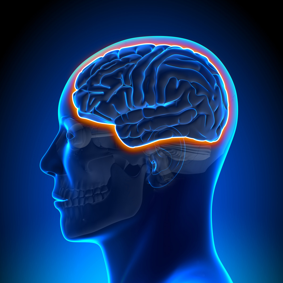
The blood-brain barrier — blood vessels with a highly specialized layer of endothelial cells tightly glued together by complexes called tight junctions — protects not only the brain but also the spine from getting into contact with potentially harmful chemicals, microbes and even cells that circulate in the blood.
“In autoimmune diseases like multiple sclerosis, immune cells that enter the brain and spinal cord cause disease,” Sarah Lutz, the study’s co-lead author, said in a news release. “A better understanding of how these cells cross the blood-brain barrier will aid our efforts to develop specific therapies to keep them out.”
Along with other researchers, Lutz, an assistant professor of anatomy and cell biology at the University of Illinois at Chicago College of Medicine, used a mouse model of human MS — the so-called experimental autoimmune encephalomyelitis (EAE) model — to investigate how Th1 and Th17 immune cells gain access to neurons.
These mice, as well as healthy control mice, were made to express a fluorescent protein of the endothelial cells present in tight junctions. Researchers performed imaging of this protein in real-time.
Their analysis showed that in the presence of Th17 cells, the tight junctions that keep the blood-brain barrier whole were severely damaged in EAE mice. This deterioration also happened early in the disease course.
Within three days, Th1 cells entered the animals’ brains and began degrading myelin and neurons. But, unlike Th17 cells that accessed the brain via the tight junctions, the Th1 cells used structures called caveolae that exist at the surface of endothelial cells.
Caveolae are invaginations of the cells’ membrane that create small pits and are a way for cells to easily exchange molecules. In fact, EAE mice that were genetically engineered to not express caveolae had almost no Th1 cells in the brain and spine.
“This is the first time we have ever seen, in live animals in real time, the different means by which these two cell types gain access to myelin and nerves,” said Lutz. “Now that we know how these cells get to neurons, drugs or small molecules can be designed that interfere with or block each of these processes to help treat and possibly prevent multiple sclerosis.”

michele garneau
i use choline a type of b vitamin to prevent them from crossing the blood barrier .this way my brain hasnt formed any new lesions in 2 yrs .. thank you m. garneau
Mark Hill
Thanks for this article. It's exactly the right approach to preventing MS. Very rare and important research, well done Sarah Lutz. As a progressive MS patient I had given up hope that any sensible research was happening. This is the first sign that anyone is doing something which might work. It may not happen in my lifetime but there is hope here that one day this wretched illness might be prevented.