
Decline in Brain Volume Driven by Gray Matter Atrophy in MS Patients Despite Tysabri Treatment
Written by |

The progressive decline in brain volume in multiple sclerosis (MS) patients, despite treatment with the disease-modifying therapy Tysabri (natalizumab), is driven by atrophy — shrinkage due to the degeneration of cells — in gray matter and not white matter structures, a new study reports.
This finding points to new markers for MS disease progression, and the need to monitor the effectiveness of treatment using magnetic resonance imaging (MRI).
The research article, Gray matter atrophy in multiple sclerosis despite clinical and lesion stability during natalizumab treatment,” was published in the journal PLOS ONE.
Brain volume loss is an important marker for assessing atrophy and disability in MS patients. However, the contribution of gray and white matter to whole brain volume loss in MS warrants further examination. Additionally, there is a need to identify markers to assess the effectiveness of known disease-modifying treatments so that MS patients can receive optimal care.
Join the MS forums: an online community especially for patients with MS.
Brain atrophy measurements, both global and regional (whole and partial), assess net tissue damage, reflecting the sum of demyelination — damage to the protective coat (called myelin) that surrounds nerve fibers — and injury and loss of brain cells. Global brain atrophy has predictive importance for MS disease progression, while regional brain atrophy is a more sensitive and reliable indicator of disease status.
Tysabri, marketed by Biogen, is a monoclonal antibody that has potent anti-inflammatory effects, and is able to reduce relapses and disability progression in patients with relapsing-remitting MS (RRMS).
In the pivotal Phase 3 trial AFFIRM in the early 2000s, Tysabri was shown to reduce brain atrophy only in the second year of treatment. Some researchers have since suggested that Tysabri may be ineffective in its first year of administration due to pseudoatrophy — a rapid, short-term drop in brain volume over a few months when patients start an anti-inflammatory therapy.
Now, a team led by researchers from The University of Chicago and the University of Turku, Finland, conducted a retrospective study to examine global and regional white and gray matter atrophy, to identify MRI-based markers for MS disease progression, and assess the effectiveness of Tysabri treatment in 20 clinically stable RRMS patients. The patients’ median age 39.5 years and were treated with the therapy for two to five years.
Clinical assessments were performed every four to six months, in which patients were assessed for brain lesions by MRI, and for their score on the Expanded Disability Status Scale (EDSS), which is a method to quantify disability in MS. Quantitative assessment of T1 hypointense lesions over time also was done using a new technique, called the T1 relaxation value, that reflects edema, demyelination, and axonal loss.
Results showed that although patients did not worsen physically and mentally with Tysabri treatment in general (all patients remained clinically stable), there was a substantial median brain volume loss of 2 percent over an average period of three years. This brain volume loss could not be explained by brain lesions, pseudoatrophy, or EDSS score.
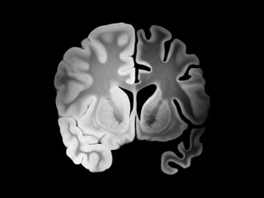
The researchers did observe a decline in gray matter, but not in white matter structures. Specifically, a brain region called thalamus (largely gray matter) demonstrated volume loss over time, but the corpus callosum (a white matter structure) did not.
Concerning MRI lesions, the team assessed specific T1 hypointense lesions (areas of relatively severe damage). Results showed a decrease in T1 values in both gray matter and white matter lesions, but the changes were not significant.
Overall, the study shows that although Tysabri could seemingly halt disease progression in white matter, gray matter atrophy may not be amenable to treatment.
“These results demonstrate that brain volume loss in MS is primarily driven by gray matter changes, and may be independent of clinically effective treatment,” such as Tysabri, the researchers concluded.
The team emphasized that “clinical trials with longer periods of observation are needed to assess many of these deficiencies, and must include separate examination of gray and white matter.”


Ant
Didn't the same story appear a week or so ago?